RMS Foundation
Robert Mathys-Strasse 1
2544 Bettlach
Schweiz
Tel. +41 32 644 2000


Unsere Forschung richtet sich neben weiteren Fachgebieten vor allem auf den menschlichen Bewegungsapparat mit Fokus auf Geweberegeneration und Implantaten. Zusätzlich fokussieren wir uns auf die Herstellung, Bearbeitung und Charakterisierung von Werkstoffen und Oberflächen. Die Resultate aus unserer Forschungstätigkeit publizieren wir in international anerkannten Fachzeitschriften.
Hier finden Sie die Auflistung von Publikationen mit Beteiligung der RMS Foundation nach Jahr ihrer Erscheinung.
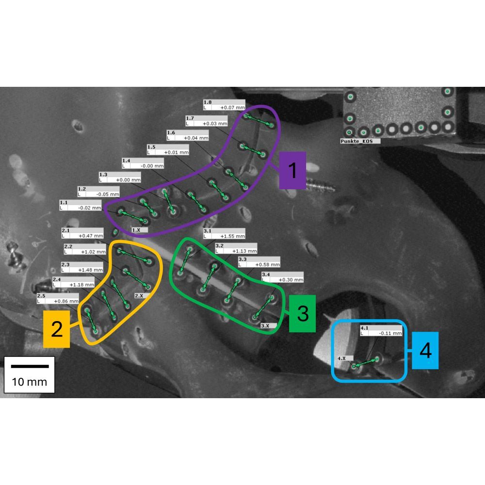
Background/Objectives: Managing acetabular fractures remains a surgical challenge, particularly in cases involving traumatic pelvic discontinuity (PD). The optimal method for achieving primary stability is unclear, and biomechanical evidence comparing established techniques is limited. The goal of this biomechanical study is to evaluate if a Ganz reinforcement ring with the addition of a posterior-column plate and anterior-column screw (GRP) provides stability comparable to a Burch-Schneider reinforcement ring (BSR) with an additional anterior- and posterior-column screws construct.
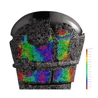
Methods: The primary biomechanical stability of two acetabular “fix-and-replace” techniques—BSR versus GRP—using standardized 4th-generation Sawbones® hemipelvis models with T-type fractures (PD) was compared. Relative 3D micromotions at the fracture site (Zone 1: Posterior-column; Zone 2:
Anterior-column; Zone 3: Oblique to transverse fracture, and Zone 4: Ischiopubic ramus) were measured under increasing cyclic loading (100 cycles per load level) at 200 N, 400 N, 800 N, and 1200 N using an optical motion tracking system. A detected fracture gap of 1000 µm or more during/after the cyclic load was defined as fixation failure.
Results: Fixation failure was not observed in any of the six artificial hemipelves with treated (3 BSR, 3 GRP) T-type acetabular fractures. Under cyclic, increasing load (200–1200 N), the mean fracture gap remained small at 200 N and 400 N with no significant differences between techniques. At 800 N, GRP fixation showed a non-significant increase in micromotion. At 1200 N, significantly greater displacements were observed in Zones 2–4 with GRP compared to BSR (p < 0.005), whereas no difference was found in Zone 1 (p = 0.424). Modelled slope and intercept comparisons confirmed a significantly steeper increase in fracture gap with GRP in zones 2–4 at higher loads (≥800 N, p < 0.01) while remaining under 1000 µm.
Conclusions: Both fixation methods demonstrated sufficient construct stability without catastrophic failure, with minimal displacement (<1 mm) and with no significant difference in stability at the posterior column.
Aims
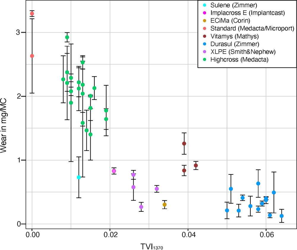
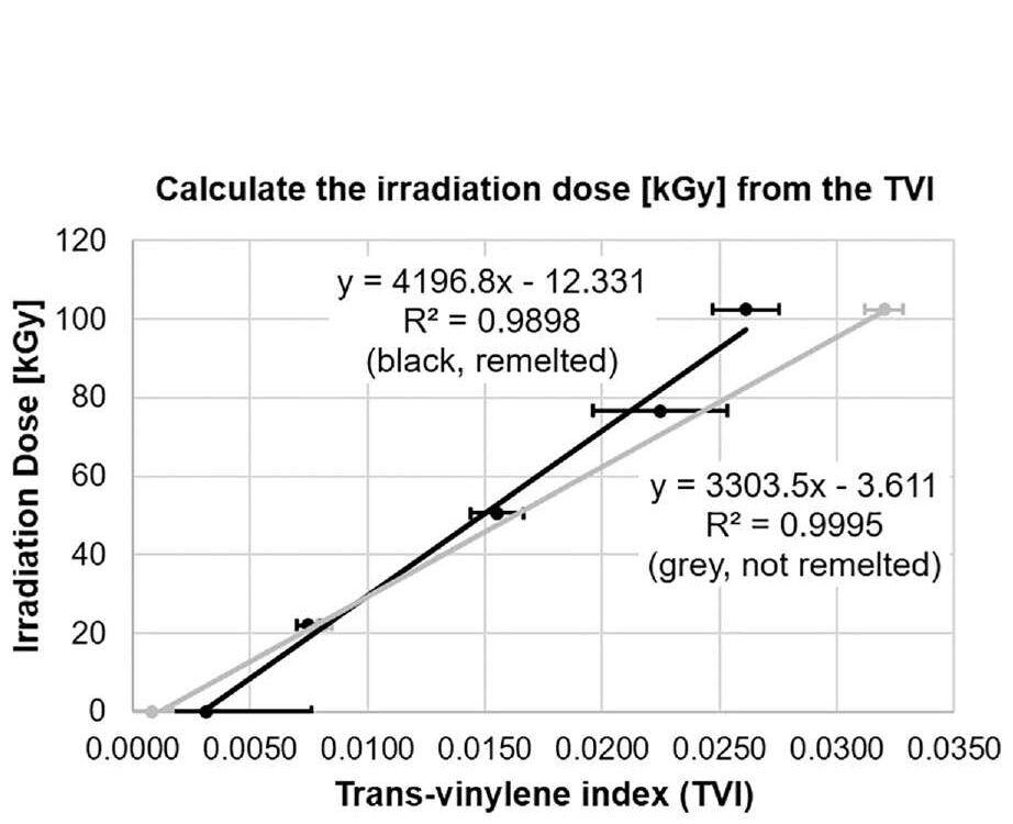
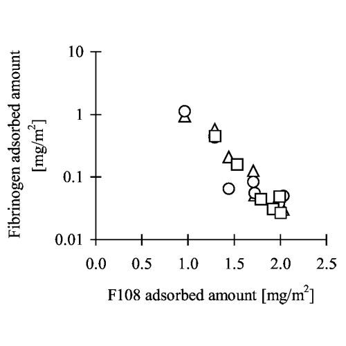
Highly cross-linked polyethylene (HXLPE) greatly reduces wear in total hip arthroplasty, compared to conventional polyethylene (CPE). Cross-linking is commonly achieved by irradiation. This study aimed to compare the degree of cross-linking and in vitro wear rates across a cohort of retrieved and unused polyethylene cups/liners from various brands.
Methods
Polyethylene acetabular cups/liners were collected at one centre from 1 April 2021 to 30 April 2022. The trans-vinylene index (TVI) and oxidation index (OI) were determined by Fourier-transform infrared spectrometry. Wear was measured using a pin-on-disk test.
Results
A total of 47 specimens from ten brands were included. The TVI was independent of time in vivo. A linear correlation (R2 = 0.995) was observed between the old and current TVI standards, except for vitamin E-containing polyethylene. The absorbed irradiation dose calculated from the TVI corresponded to product specifications for all but two products. For one electron beam-irradiated HXLPE, a mean dose of 241% (SD 18%) of specifications was determined. For another, gamma-irradiated HXLPE, a mean 41% (SD 13%) of specifications was determined. Lower wear was observed for higher TVI.
Conclusion
The TVI is a reliable measure of the absorbed irradiation dose and does not alter over time in vivo. The products of various brands differ by manufacturing details and consequently cross-linking characteristics. Absorption and penetration of electron radiation and gamma radiation differ, potentially leading to higher degrees of cross-linking for electron radiation. There is a non-linear, inverse correlation between TVI and in vitro wear. The wear resistance of the HXLPE with low TVI was reduced and more comparable to CPE.
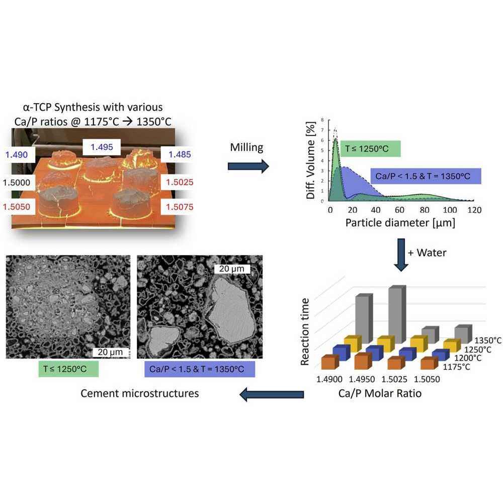
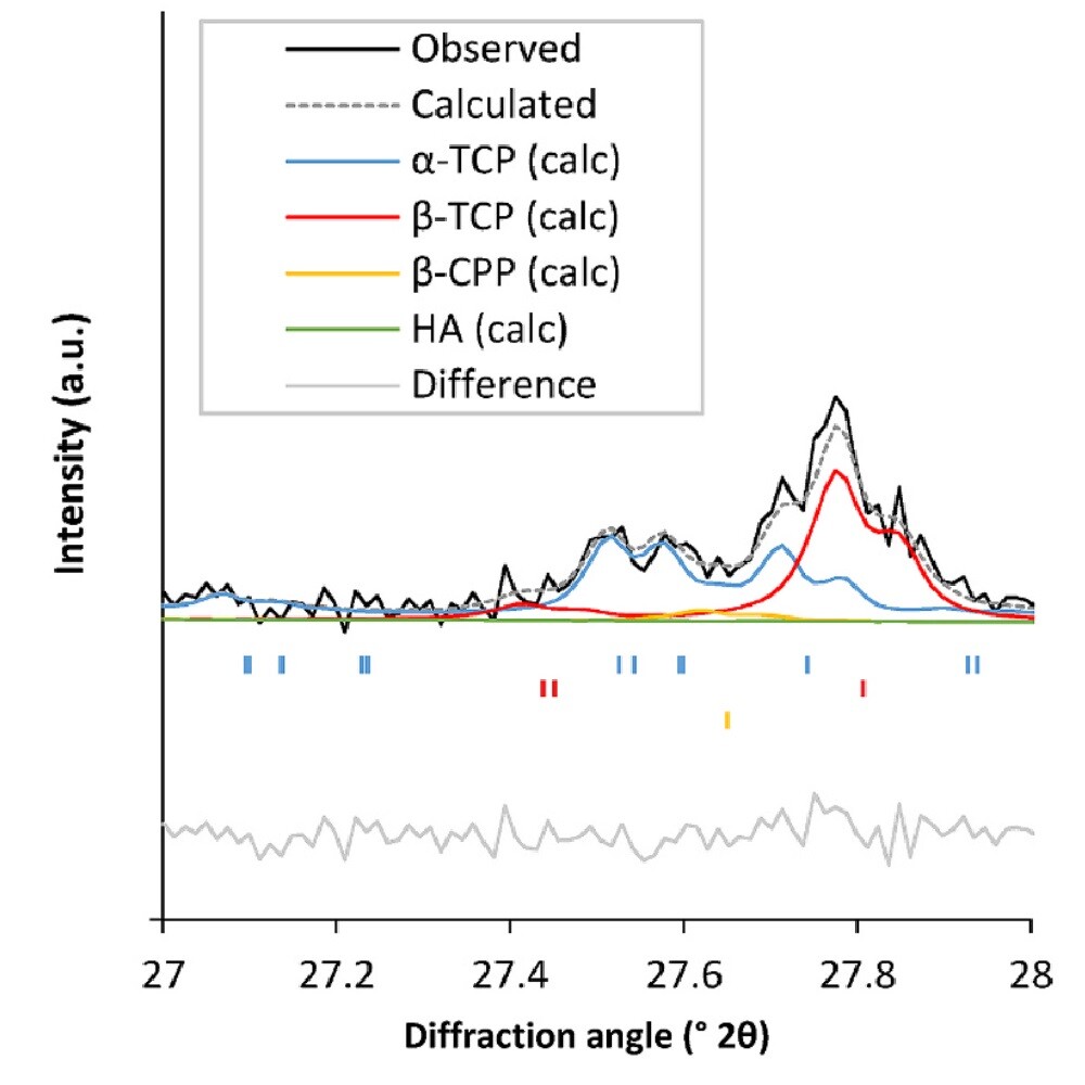
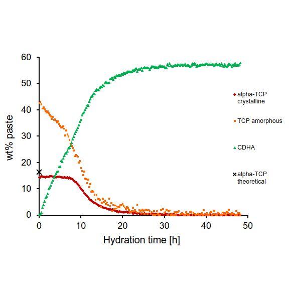
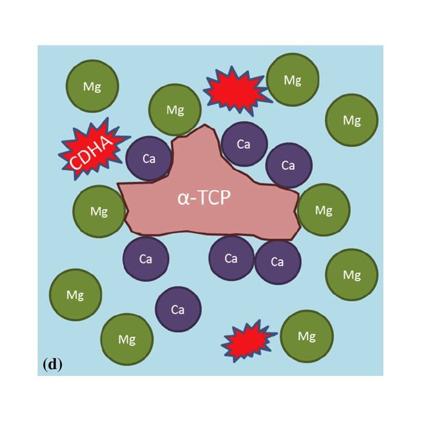
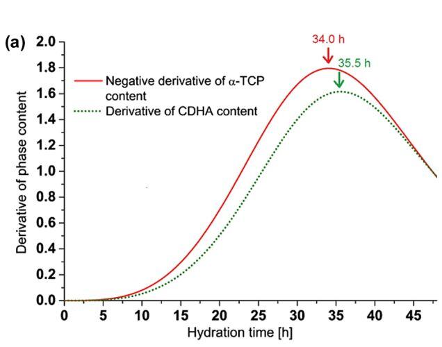
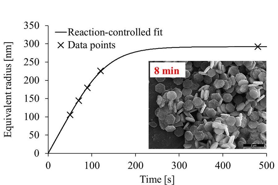
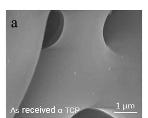
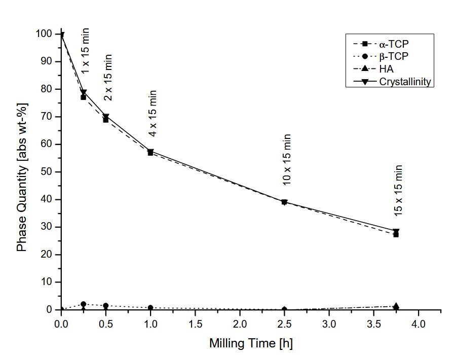
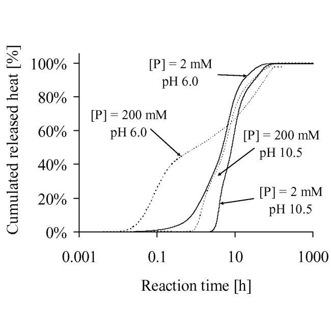
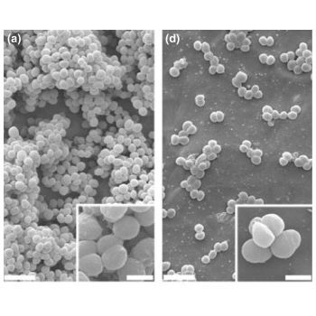
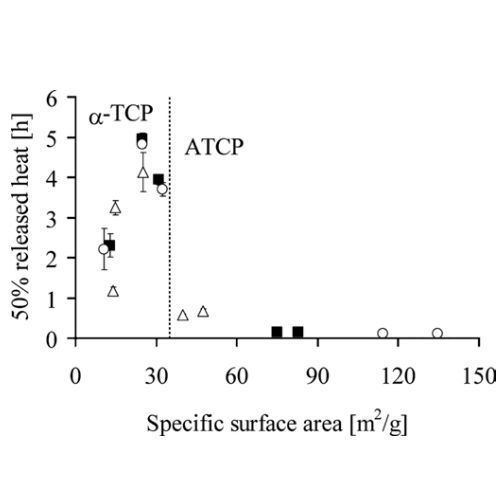
α-tricalcium phosphate (α-TCP) is the most widespread raw material for hydraulic calcium phosphate cements (CPCs). CPCs are widely used in bone repair due to their injectability, setting ability, and osteoconductivity. This study investigated the reactivity of α-TCP powders, focusing on the impact of minor phase impurities, β-calcium pyrophosphate and hydroxyapatite, and the synthesis temperature. The α-TCP powders were synthesized via a solid-state reaction of calcium carbonate and anhydrous dicalcium phosphate, with varying Ca/P molar ratios (1.4850–1.5075) and synthesis temperatures (1175°C–1350 °C). Powders produced with a Ca/P molar ratio below 1.50 and synthesized at a temperature above the melting point of β-CPP (1296 °C) had a broader size distribution and a two to fourfold lower hydraulic reactivity. Conversely, a higher Ca/P molar ratio improved reactivity. The study underscores the importance of precise control over synthesis parameters to enhance the performance of α-TCP-based CPCs, offering insights for optimizing material design in biomedical applications.
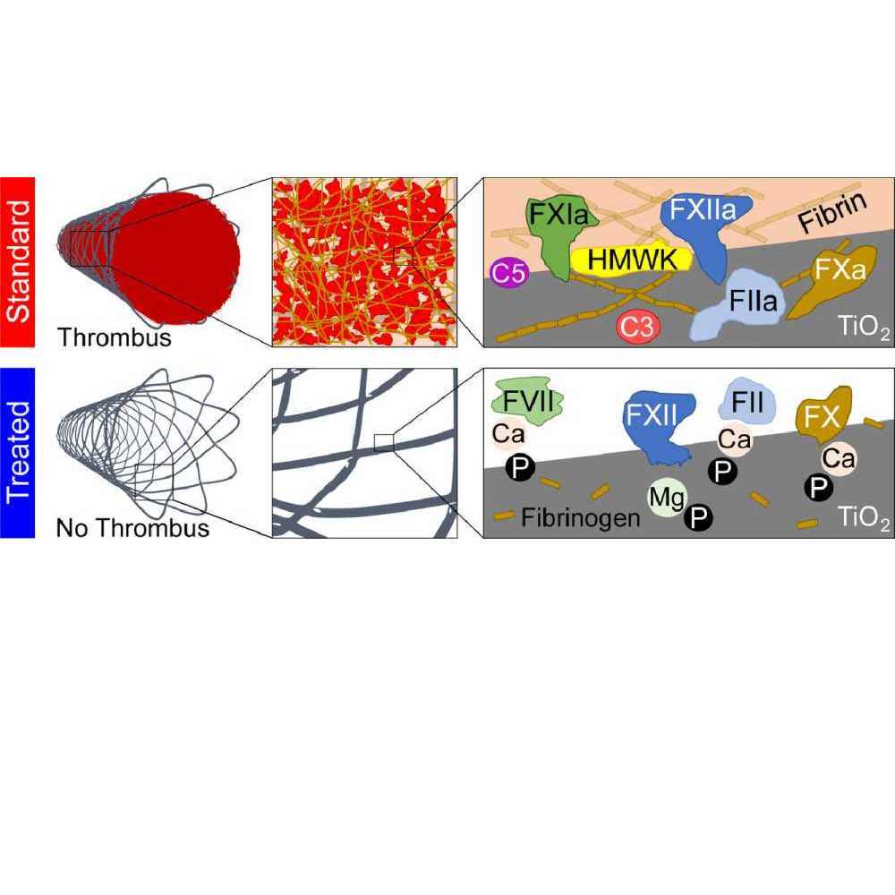
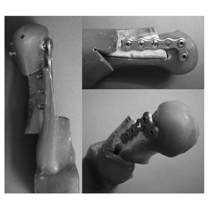
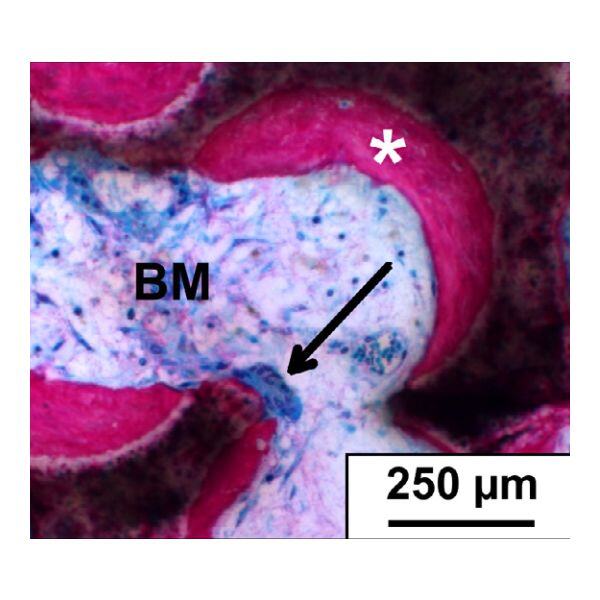
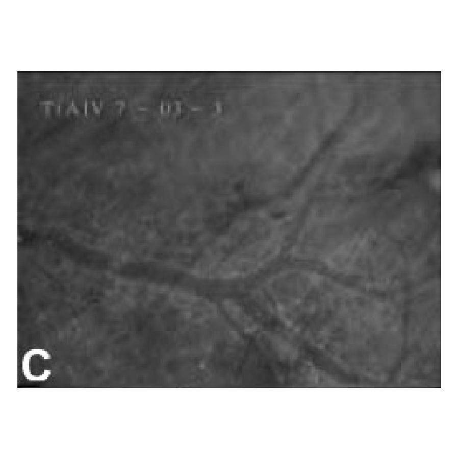
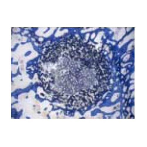
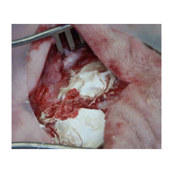
Titanium as the leading implant material in locked plating is challenged by polymers such as carbon fiber-reinforced polyetheretherketone (CFR-PEEK), which became the focus of interest of researchers and manufacturers in recent years. However, data on human tissue response to these new implant materials are rare.
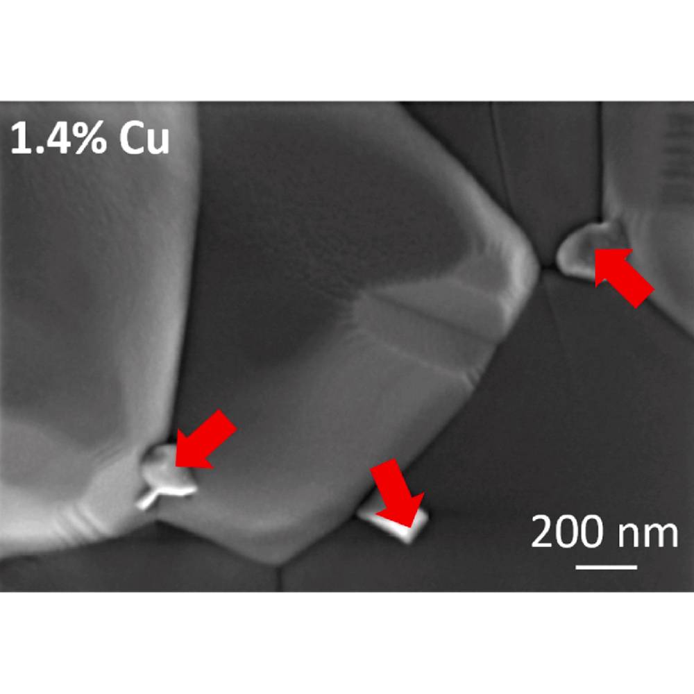
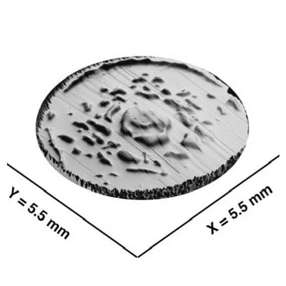
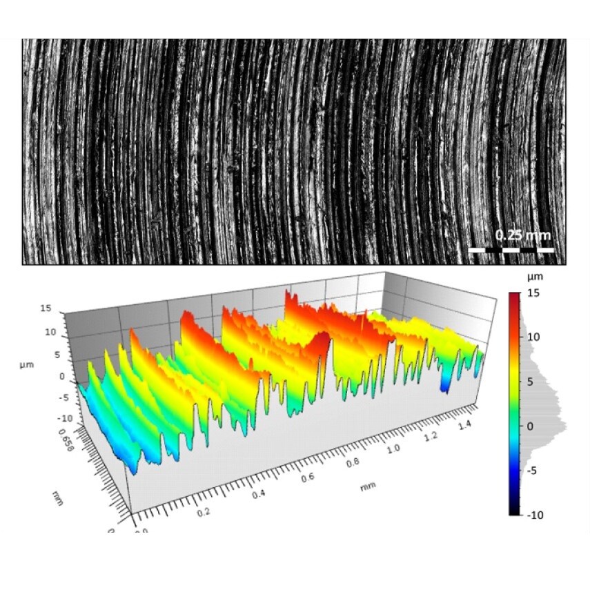
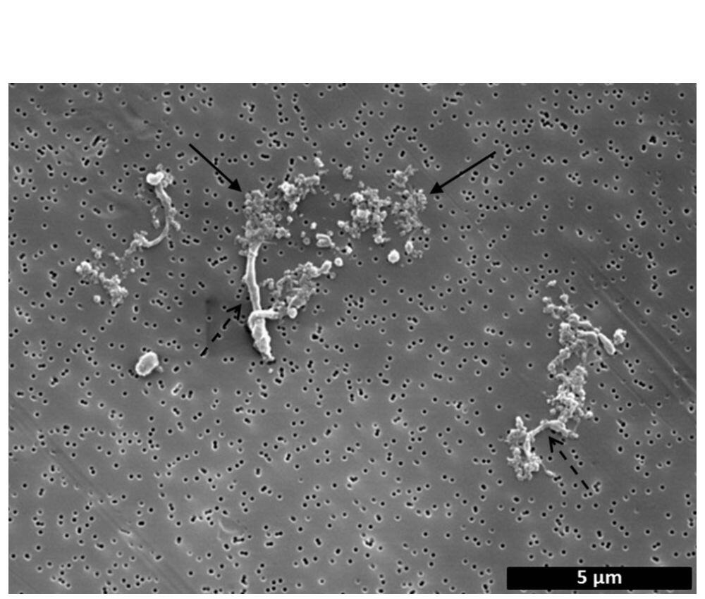
Osteosynthesis plates and peri–implant soft tissue samples of 16 healed proximal humerus fractures were examined ( n = 8 CFR-PEEK, n = 8 titanium). Soft tissue was analyzed by immunohistochemistry and μCT. The entrapped foreign bodies were further examined for their material composition by FTIR. To gain insight into their origin and formation mechanism, explanted and new plates were evaluated by SEM, EDX, profilometry and HR-CT.
In the peri–implant soft tissue of the CFR-PEEK plates, an inflammatory tissue reaction was detected. Tissues contained foreign bodies, which could be identified as tantalum wires, carbon fiber fragments and PEEK particles. Titanium particles were also found in the peri–implant soft tissue of the titanium plates but showed a less intense surrounding tissue inflammation in immunohistochemistry. The surface of explanted CFR-PEEK plates was rougher and showed exposed and broken carbon fibers as well as pro- truding and deformed tantalum wires, especially in used screw holes, whereas scratches were identified on the titanium plate surfaces.
Particles were present in the peri–implant soft tissue neighboring both implant materials and could be clearly assigned to the plate material. Particles from both plate materials caused detectable tissue inflammation, with more inflammatory cells found in soft tissue over CFR-PEEK plates than over titanium plates.
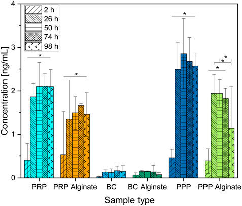
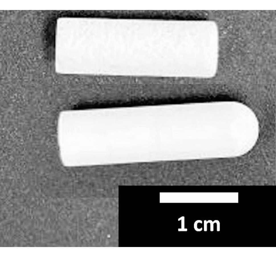
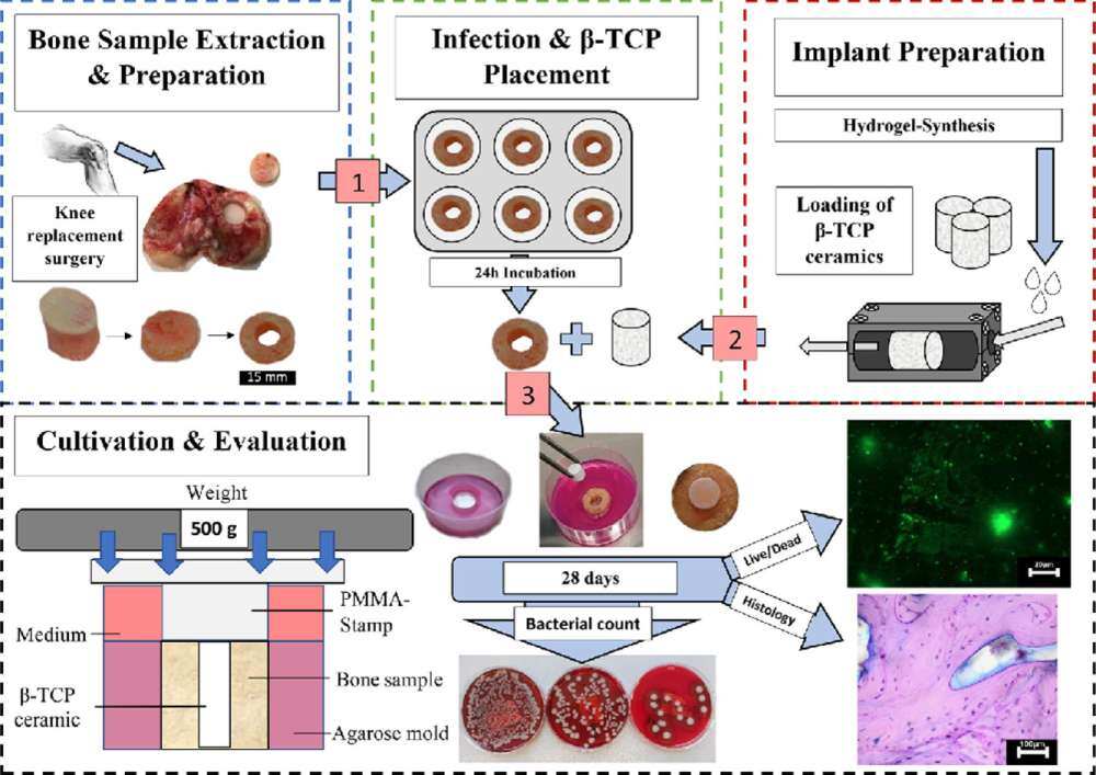
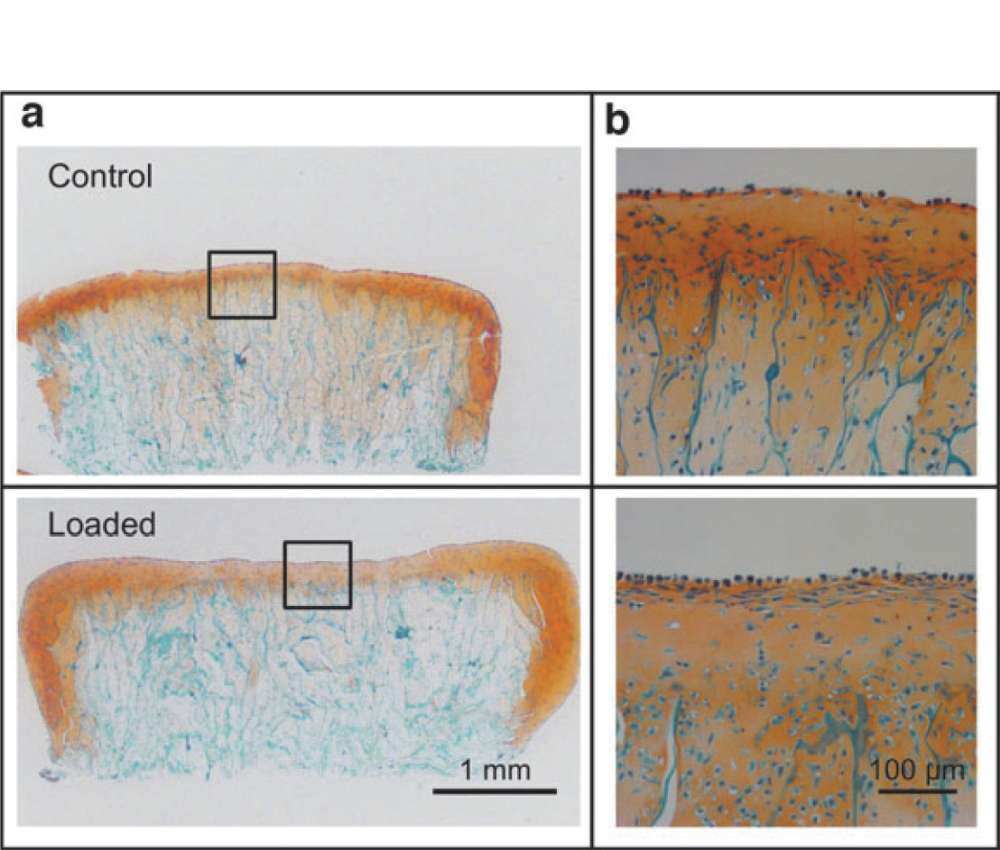
Introduction: β-TCP ceramics are bone replacement materials that have recently been tested as a drug delivery system that can potentially be applied to endogenous substances like growth factors found in blood platelets to facilitate positive attributes.
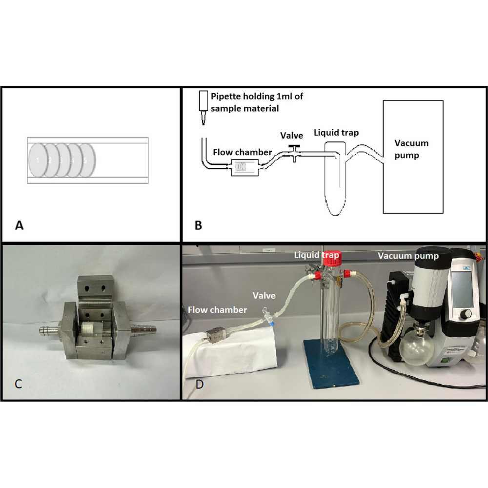
Methods: In this work, we used flow chamber loading to load β-TCP dowels with blood suspensions of platelet-rich plasma (PRP), platelet-poor plasma (PPP), or buffy coat (BC) character. PRP and BC platelet counts were adjusted to the same level by dilution. Concentrations of TGF-β1, PDGF-AB, and IGF-1 from dowel-surrounding culture medium were subsequently determined using ELISA over 5 days. The influence of alginate was additionally tested to modify the release.
Results: Concentrations of TGF-β1 and PDGF-AB increased and conclusively showed a release from platelets in PRP and BC compared to PPP. The alginate coating reduced the PDGF-AB release but did not reduce TGF-β1 and instead even increased TGF-β1 in the BC samples. IGF-1 concentrations were highest in PPP, suggesting circulating levels rather than platelet release as the driving factor. Alginate samples tended to have lower IGF-1 concentrations, but the difference was not shown to be significant.
Discussion: The release of growth factors from different blood suspensions was successfully demonstrated for β-TCP as a drug delivery system with release patterns that correspond to PRP activation after Ca2+-triggered activation. The release pattern was partially modified by alginate coating.
Objectives
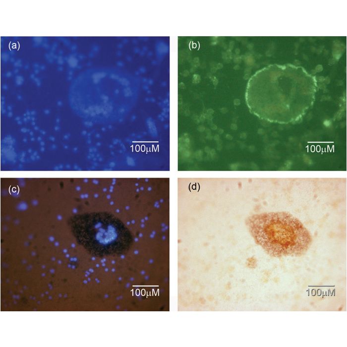
Administration of gadolinium-based contrast agents (GBCA) in magnetic resonance imaging results in the long-term retention of gadolinium (Gd) in tissues and organs, including the bone, and may affect their function and metabolism. This study aims to investigate the effects of Gd and GBCA on the proliferation/survival, differentiation, and function of bone cell lineages.
Materials and Methods
Primary murine osteoblasts (OB) and osteoclast progenitor cells (OPC) isolated from C57BL/6J mice were used to test the effects of Gd3+ (12.5–100 μM) and GBCA (100–2000 μM). Cultures were supplemented with the nonionic linear Gd-DTPA-BMA (gadodiamide), ionic linear Gd-DTPA (gadopentetic acid), and macrocyclic Gd-DOTA (gadoteric acid). Cell viability and differentiation were analyzed on days 4–6 of the culture. To assess the resorptive activity of osteoclasts, the cells were grown in OPC cultures and were seeded onto layers of amorphous calcium phosphate with incorporated Gd.
Results
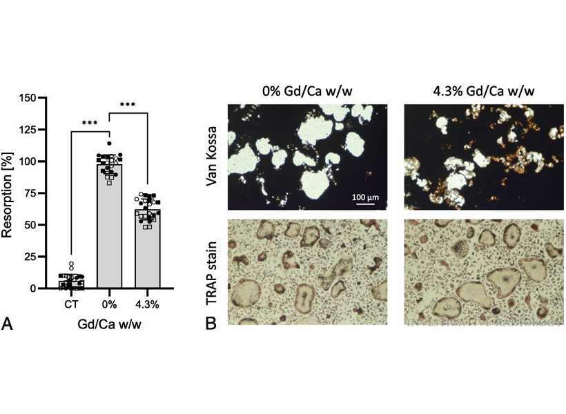
Gd3+ did not affect OB viability, but differentiation was reduced dose-dependently up to 72.4% ± 6.2%–73.0% ± 13.2% (average ± SD) at 100 μM Gd3+ on days 4–6 of culture as compared with unexposed controls (P < 0.001). Exposure to GBCA had minor effects on OB viability with a dose-dependent reduction up to 23.3% ± 10.2% for Gd-DTPA-BMA at 2000 μM on day 5 (P < 0.001). In contrast, all 3 GBCA caused a dose-dependent reduction of differentiation up to 88.3% ± 5.2% for Gd-DTPA-BMA, 49.8% ± 16.0% for Gd-DTPA, and 23.1% ± 8.7% for Gd-DOTA at 2000 μM on day 5 (P < 0.001). In cultures of OPC, cell viability was not affected by Gd3+, whereas differentiation was decreased by 45.3% ± 9.8%–48.5% ± 15.8% at 100 μM Gd3+ on days 4–6 (P < 0.05). Exposure of OPC to GBCA resulted in a dose-dependent increase in cell viability of up to 34.1% ± 11.4% at 2000 μM on day 5 of culture (P < 0.001). However, differentiation of OPC cultures was reduced on day 5 by 24.2% ± 9.4% for Gd-DTPA-BMA, 47.1% ± 14.0% for Gd-DTPA, and 38.2% ± 10.0% for Gd-DOTA (P < 0.001). The dissolution of amorphous calcium phosphate by mature osteoclasts was reduced by 36.3% ± 5.3% upon incorporation of 4.3% Gd/Ca wt/wt (P < 0.001).
Conclusions
Gadolinium and GBCA inhibit differentiation and activity of bone cell lineages in vitro. Thus, Gd retention in bone tissue could potentially impair the physiological regulation of bone turnover on a cellular level, leading to pathological changes in bone metabolism.
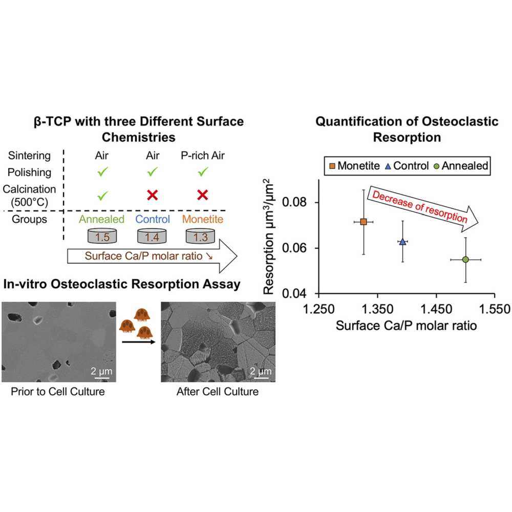
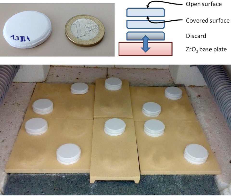
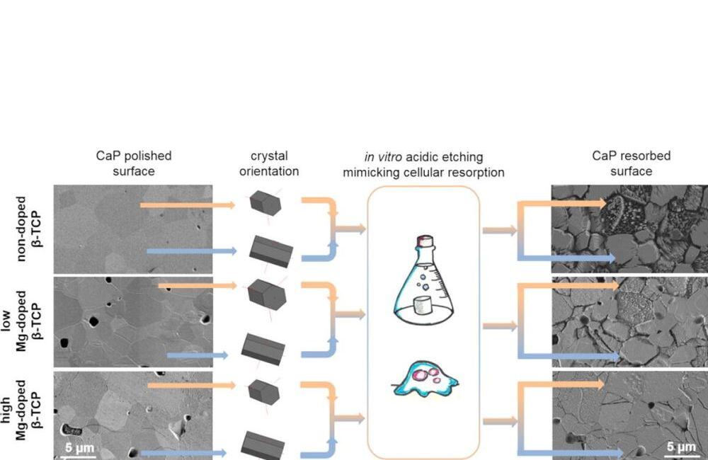
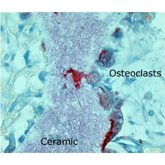
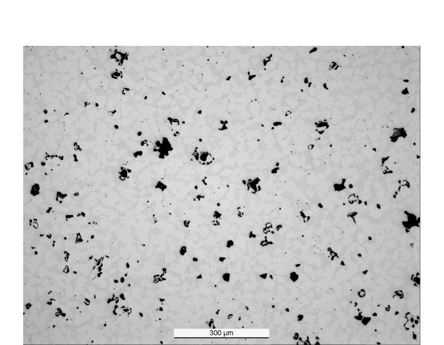
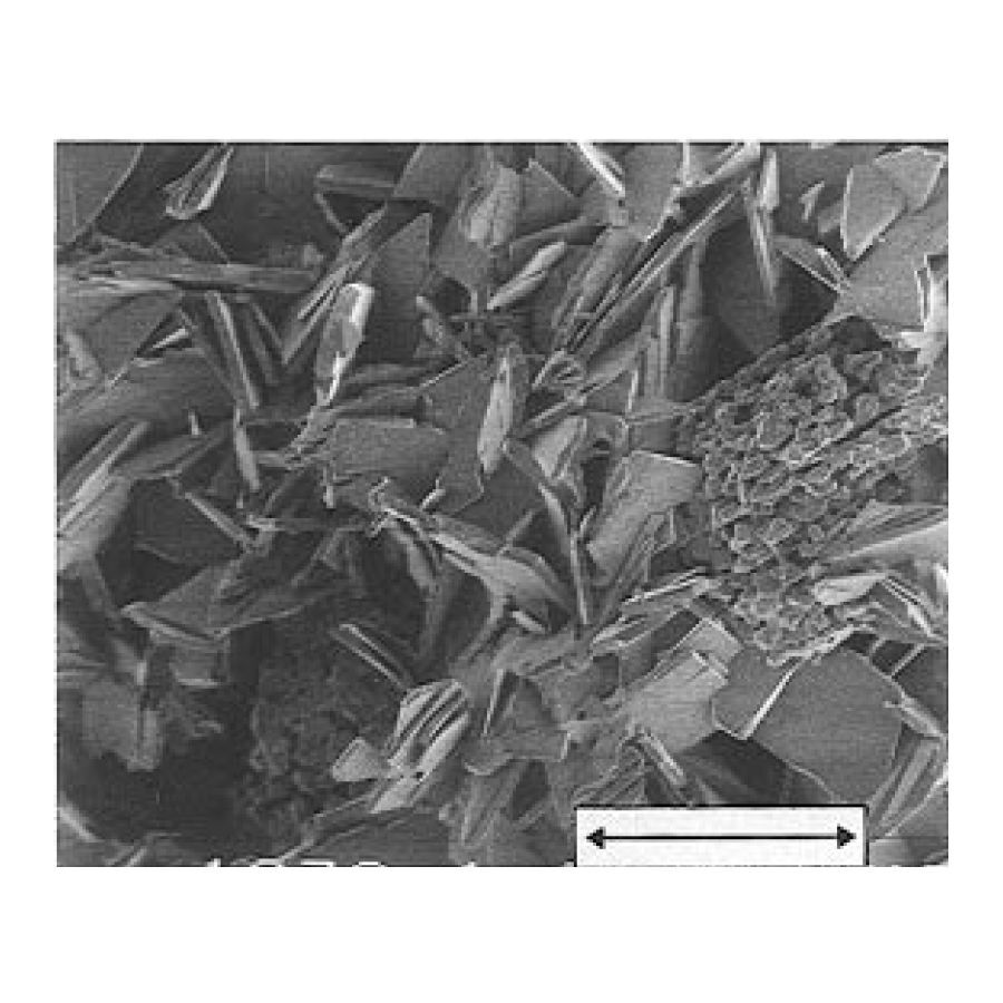
One of the most widely used materials for bone graft substitution is β‐Tricalcium phosphate (β‐TCP; β-Ca3(PO4)2). β-TCP is typically produced by sintering in air or vacuum. During this process, evaporation of phosphorus (P) species occurs, leading to the formation of a calcium-rich alkaline layer. It was recently shown that the evaporation of P species could be prevented by co-sintering β-TCP with dicalcium phosphate (DCPA; CaHPO4; mineral name: monetite). The aim of this study was to see how a change of sintering atmosphere could affect the physico-chemical and biological properties of β-TCP. For this purpose, three experimental groups were considered: β‐TCP cylinders sintered in air and subsequently polished to remove the surface layer (control group); the same polished cylinders after subsequent annealing at 500 °C in air to generate a calcium-rich alkaline layer (annealed group); and finally, β‐TCP cylinders sintered in a monetite-rich atmosphere and subsequently polished (monetite group). XPS analysis confirmed that cylinders from the annealed group had a significantly higher Ca/P molar ratio at their surface than that of the control group while this ratio was significantly lower for the cylinders from the monetite group. Sintering β‐TCP in the monetite-rich atmosphere significantly reduced the grain size and increased the density. Changes of surface composition affected the activity of osteoclasts seeded onto the surfaces, since annealed β-TCP cylinders were significantly less resorbed than β-TCP cylinders sintered in the monetite-rich atmosphere. This suggests that an increase of the surface Ca/P molar ratio leads to a decrease of osteoclastic resorption.
Statement of Significance
Minimal changes of surface and bulk (< 1%) composition have major effects on the ability of osteoclasts to resorb β-tricalcium phosphate (β-TCP), one of the most widely used ceramics for bone substitution. The results presented in this study are thus important for the calcium phosphate community because (i) β-TCP may have up to 5% impurities according to ISO and ASTM standards and still be considered to be “pure β-TCP”, (ii) β-TCP surface properties are generally not considered during biocompatibility assessment and (iii) a rationale can be proposed to explain the various inconsistencies reported in the literature on the biological properties of β-TCP.
Background: There are no generally accepted guidelines for polyethylene (PE) glenoid component cementation techniques. In particular, it is not known whether the backside of a PE glenoid should be fully or partially cemented – or not cemented at all. We hypothesized that cementing techniques would have an impact on cement mantle volume and integrity, as well as biomechanical stability, measured as micromotion under cyclic loading.
Methods: To address our hypothesis, 3 different cementation techniques using a single 2-peg PE glenoid design with polyurethane foam were compared regarding (1) the quality and quantity of the cement mantle and (2) biomechanical stability after cyclic loading in vitro. Eight identically cemented glenoids per group were used. Group A underwent cement application only into the peg holes, group B received additional complete cement mantle application on the backside of the glenoid, and group C received the same treatment as group B but with additional standardized drill holes in the surface of the glenoid bone for extra cement interdigitation. All glenoids underwent cyclic edge loading by 105 cycles according to ASTM F2028-14. Before and after loading, cement mantle evaluation was performed by XtremeCT and biomechanical strength and loosening were evaluated by measuring the relative motion of the implants.
Results: The cement mantle at the back of the implant was incomplete in group A as compared with groups B and C, in which the complete PE backside was covered with a homogeneous cement mantle. The cement mantle was thickest in group C, followed by group B (P = .006) and group A (P <.001). We did not detect any breakage of the cement mantle in any of the 3 groups after testing.

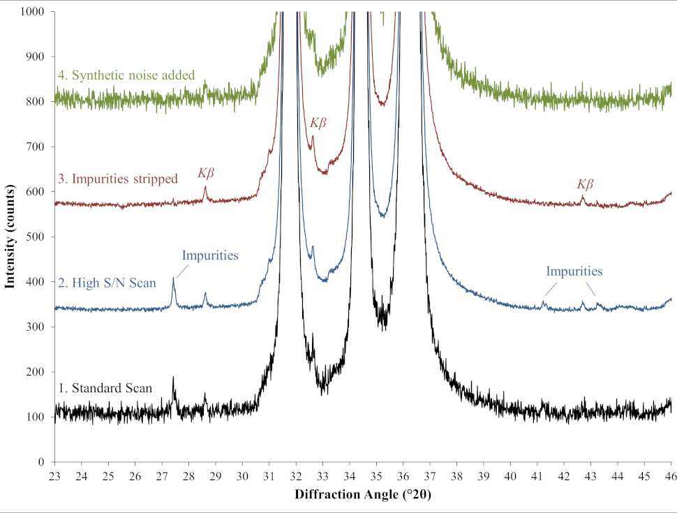
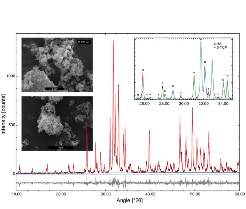
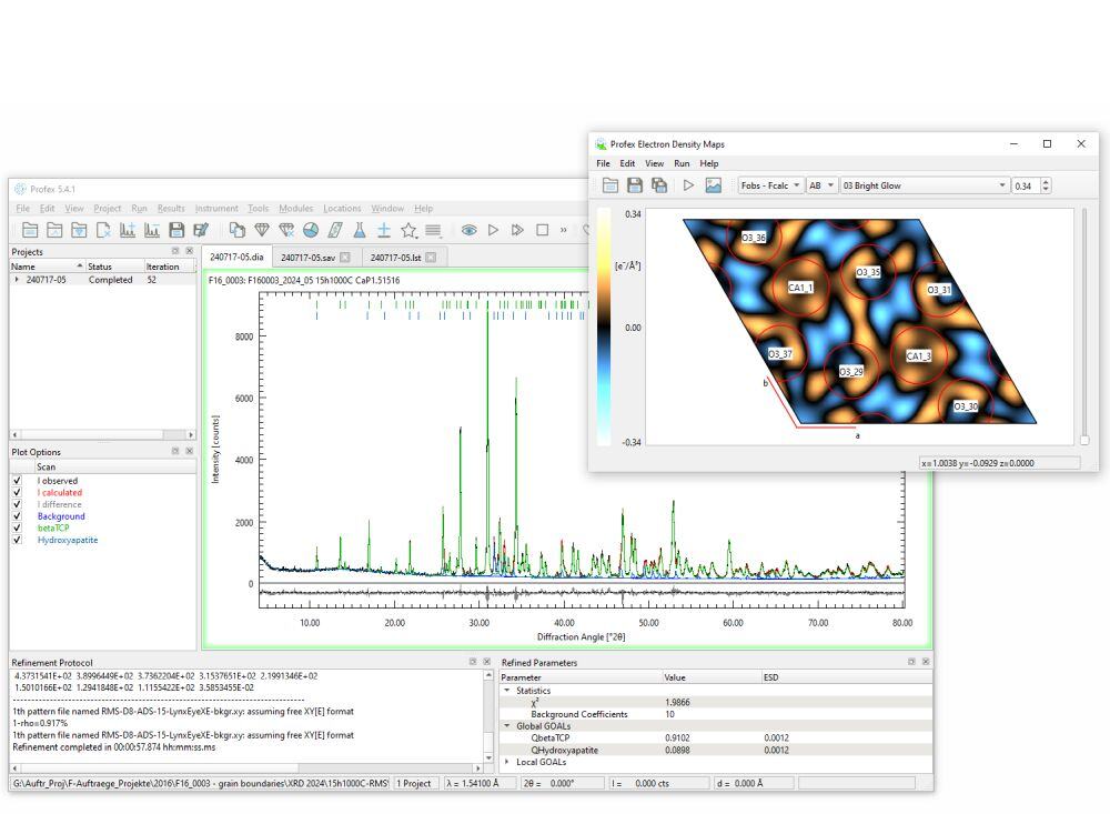
The Rietveld refinement software Profex is an open-source and platform-independent solution for the processing of powder X-ray diffraction datasets. It is based on the BGMN refinement kernel and uses a description of the diffractometer configuration to determine the instrument-related peak profile. In this article we present a Profex configuration file for the Chemistry and Mineralogy (CheMin) X-ray diffractometer (XRD), which is on-board the Mars Science Laboratory rover Curiosity. For the past decade, Curiosity has been on a mission on Mars to find out whether the planet was once habitable for microbial life. The CheMin XRD determines the mineralogical phases and abundances of Martian soil and rocks in Gale Crater, Mars. Since its arrival on Mars in 2012, Curiosity has analyzed powdered soil and rock samples with the CheMin instrument and transmitting the raw XRD data acquired back to Earth.
Adaptations of Profex to work seamlessly with CheMin XRD datasets involved creating a new configuration file for the CheMin instrument, as well as adding the Mars Mineral Compendium, a compilation of structural models specifically selected for the analysis of Mars sedimentary soil and rock samples, to Profex. Using example refinements, we demonstrate that this software solution is well suited for quantitative analysis of CheMin XRD datasets.
Titanium as the leading implant material in locked plating is challenged by polymers such as carbon fiber-reinforced polyetheretherketone (CFR-PEEK), which became the focus of interest of researchers and manufacturers in recent years. However, data on human tissue response to these new implant materials are rare.
Osteosynthesis plates and peri–implant soft tissue samples of 16 healed proximal humerus fractures were examined ( n = 8 CFR-PEEK, n = 8 titanium). Soft tissue was analyzed by immunohistochemistry and μCT. The entrapped foreign bodies were further examined for their material composition by FTIR. To gain insight into their origin and formation mechanism, explanted and new plates were evaluated by SEM, EDX, profilometry and HR-CT.
In the peri–implant soft tissue of the CFR-PEEK plates, an inflammatory tissue reaction was detected. Tissues contained foreign bodies, which could be identified as tantalum wires, carbon fiber fragments and PEEK particles. Titanium particles were also found in the peri–implant soft tissue of the titanium plates but showed a less intense surrounding tissue inflammation in immunohistochemistry. The surface of explanted CFR-PEEK plates was rougher and showed exposed and broken carbon fibers as well as pro- truding and deformed tantalum wires, especially in used screw holes, whereas scratches were identified on the titanium plate surfaces.
Particles were present in the peri–implant soft tissue neighboring both implant materials and could be clearly assigned to the plate material. Particles from both plate materials caused detectable tissue inflammation, with more inflammatory cells found in soft tissue over CFR-PEEK plates than over titanium plates.
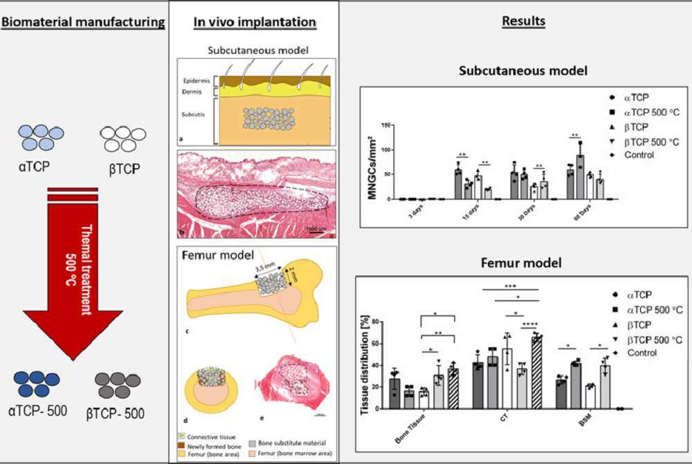
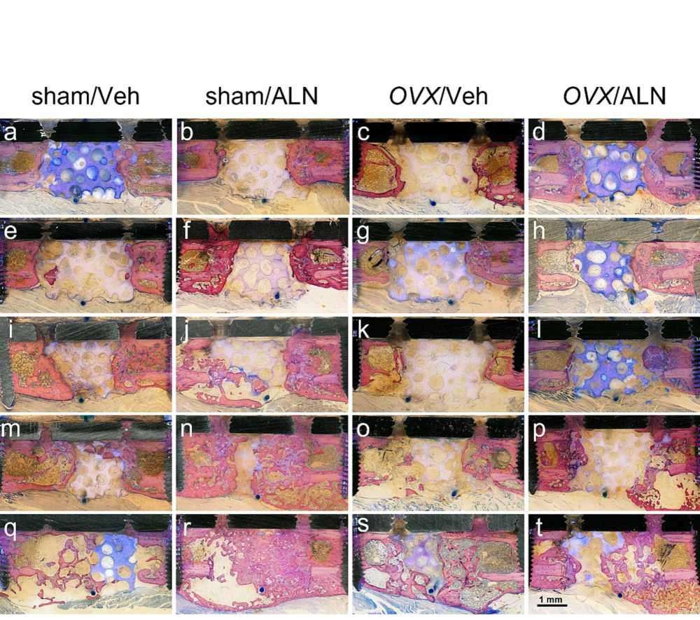
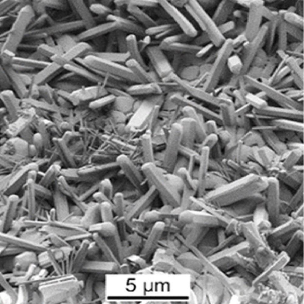
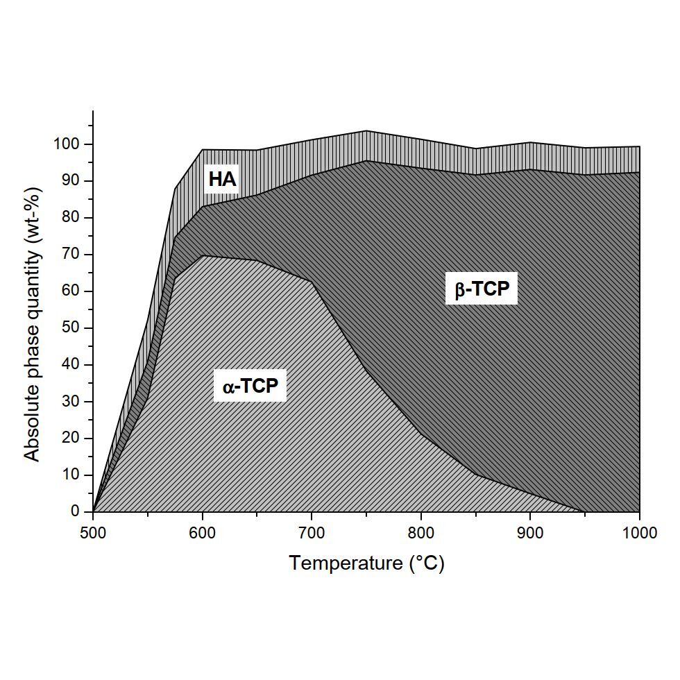
Evaporation of phosphate species during thermal treatment (> 400 °C) of calcium phosphates leads to the formation of an alkaline layer on their surface. The aim of this study was to evaluate the hypothesis that the biological response of thermally treated calcium phosphates is modified by the presence of such an alkaline layer on their surface. For this purpose, 0.125–0.180 mm α- and β-tricalcium phosphate (TCP) granules were obtained by crushing and size classification, with some being subjected to thermal treatment at 500 °C. The four types of granules (α-TCP, β-TCP, α-TCP-500 °C, and β-TCP-500 °C) were implanted subcutaneously and orthotopically in rats. Sham operations served as control.
Subcutaneously, α-TCP and β-TCP induced significantly more multinucleated giant cells (MNGCs) than calcined granules. Most of the induced MNGCs were TRAP-negative, CD-68 positive and cathepsin K-negative, reflecting a typical indication of a reaction with a foreign body. The vessel density was significantly higher in the α-TCP and β-TCP groups than it was in the α-TCP-500 °C and β-TCP-500 °C groups. In the femur model, β-TCP-500 °C induced significantly more new bone formation than that induced by β-TCP. The granule size was also significantly larger in the β-TCP-500 °C group, making it more resistant to degradation than β-TCP. The MNGC density was higher in the α-TCP and β-TCP groups than in the α-TCP-500 °C and β-TCP-500 °C groups, including cathepsin-positive, CD-68 positive, TRAP-positive and TRAP-negative MNGCs.
In conclusion, this study confirms that the biological response of calcium phosphates was affected by the presence of an alkaline layer on their surface. Thermally-treated α-TCP and β-TCP granules produced significantly fewer MNGCs and were significantly less degraded than non-thermally-treated α-TCP and β-TCP granules. Thermally treating α-TCP and β-TCP granules shifts the reaction from a foreign body reaction towards a physiological reaction by downregulating the number of induced MNGCs and enhancing degradation resistance.
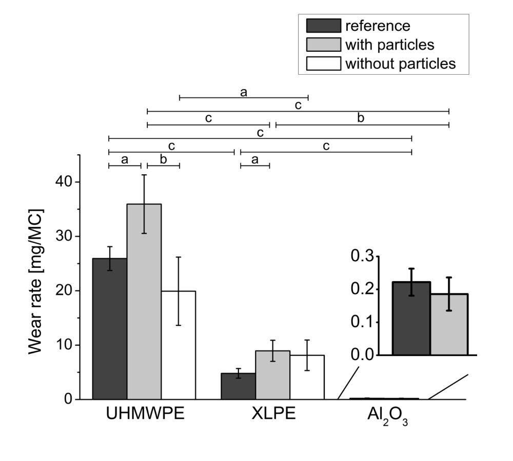
The feasibility to use poly-ether-ether-ketone (PEEK) instead of CoCrMo for femoral condyles of knee prostheses was investigated in this pin-on-disc study. This replacement would hinder the release of the toxic elements Co, Cr and Mo and this possibly at lower production costs. In this preliminary pin-on-disc study, the wear of the pairings PEEK vs. ultra-high-molecular-weight polyethylene (UHMWPE) and PEEK vs. vitamin E containing crosslinked polyethylene (XLPE) was investigated in unidirectional and multidirectional wear tests.
The articulating surfaces of the pins and discs were polished after the wear tests, while most of the initial toolmarks were removed. In addition, there were small scratches on the surfaces. Striations and protuberances were found on some of the UHMWPE samples.
All wear factors were relatively small with less than 1 × 10⁻⁶ mm3/Nm. Due to the low wear, the resulting weight loss was highly affected by the soaking of the samples. This was especially the case for the discs due to their larger surface. Thus, the wear of the discs was not used for the comparisons. The wear factors for PEEK pins and XLPE pins were in the range of (0.05 to 0.06) × 10−6 mm3/Nm, both for unidirectional and multidirectional motion. The wear of the UHMWPE pins was with (0.07 ± 0.01) × 10−6 mm3/Nm slightly higher under unidirectional motion and with (0.28 ± 0.03) × 10−6 mm3/Nm four times higher under multidirectional motion.
Thus, PEEK should be considered as an alternative to the CoCrMo alloy in knee prostheses, especially when used in combination with XLPE liners.
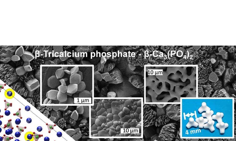
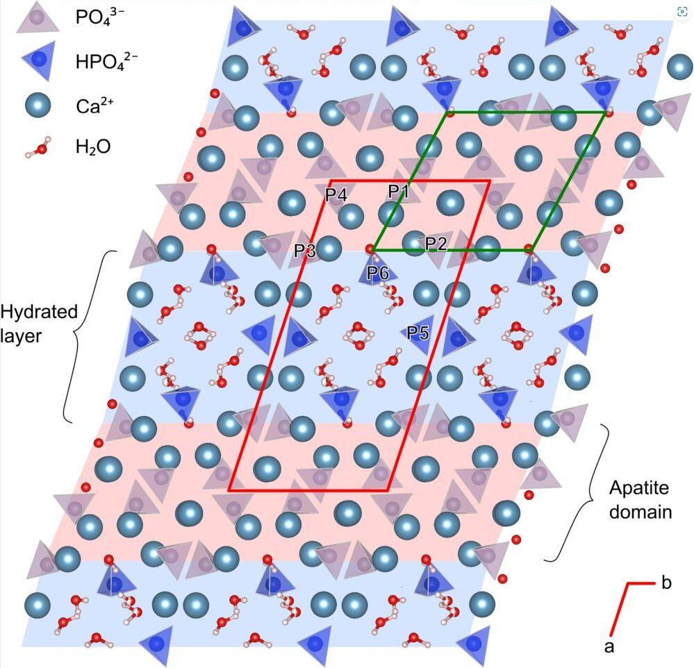
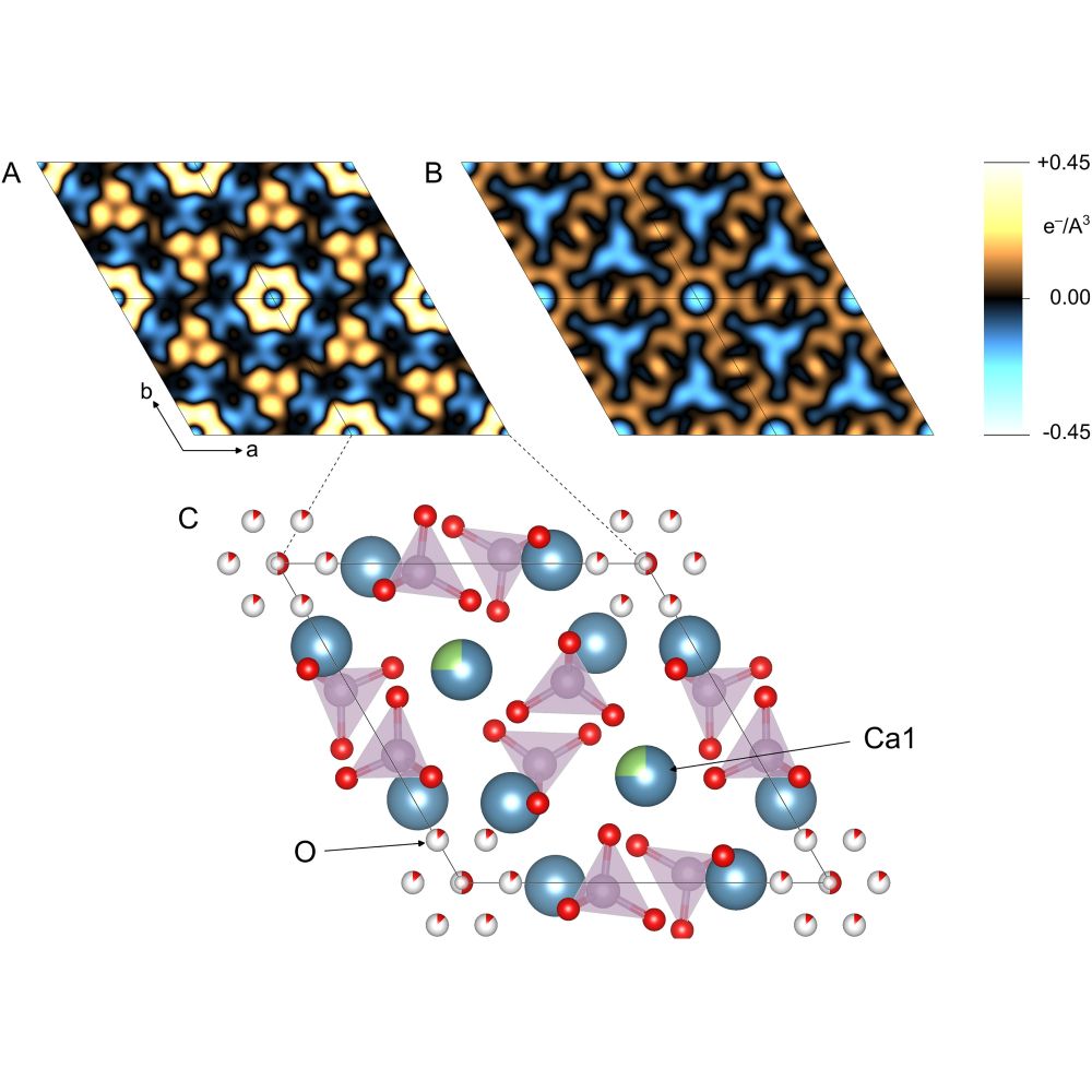
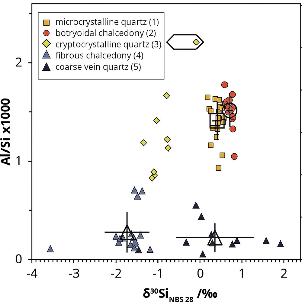
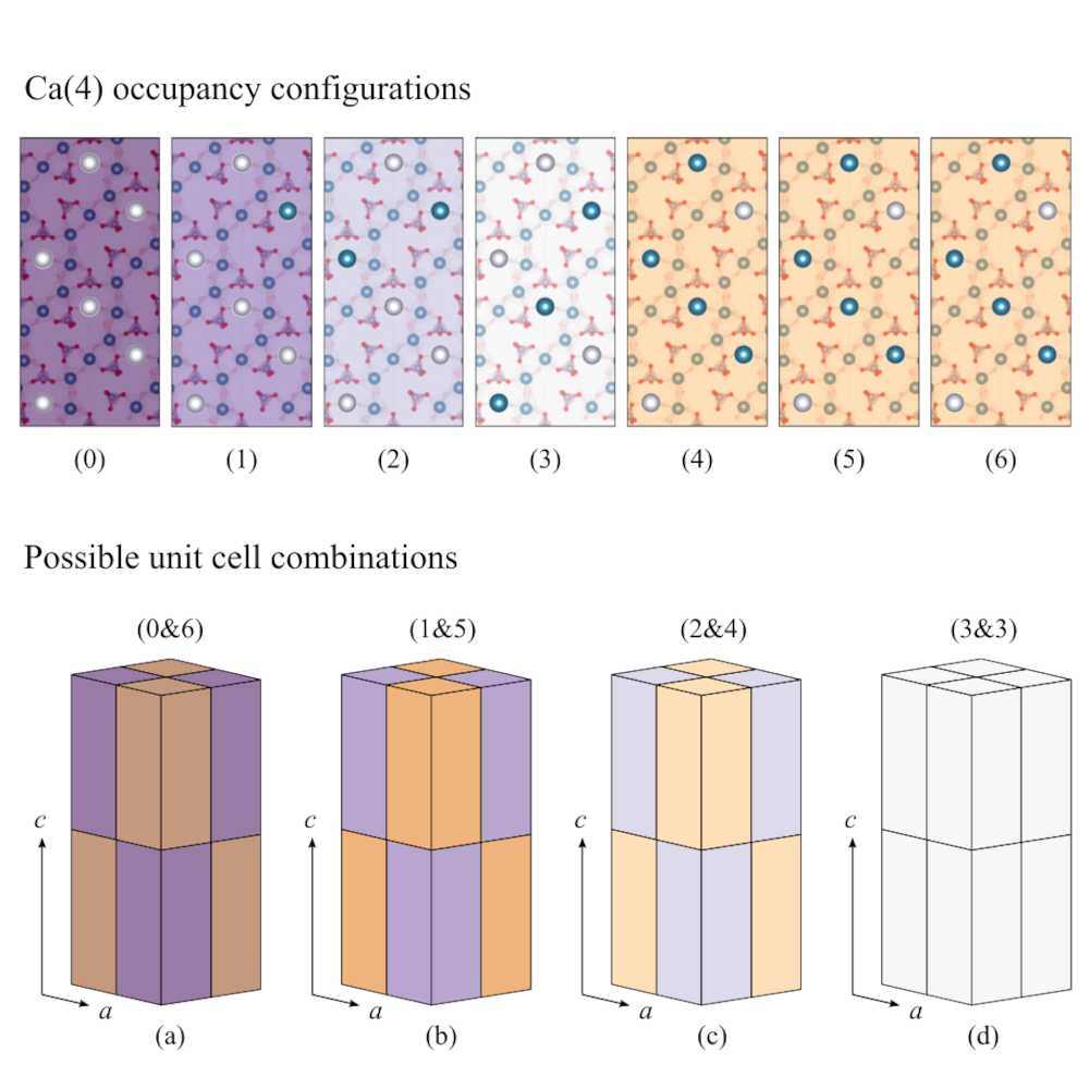
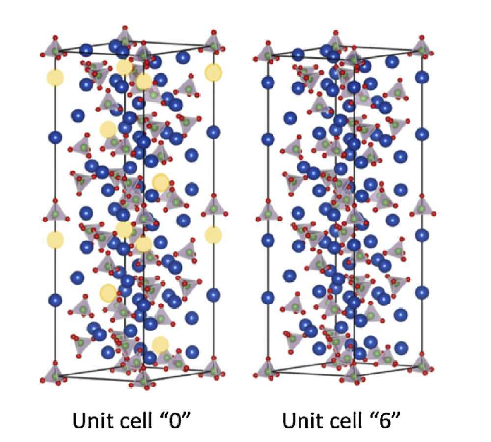
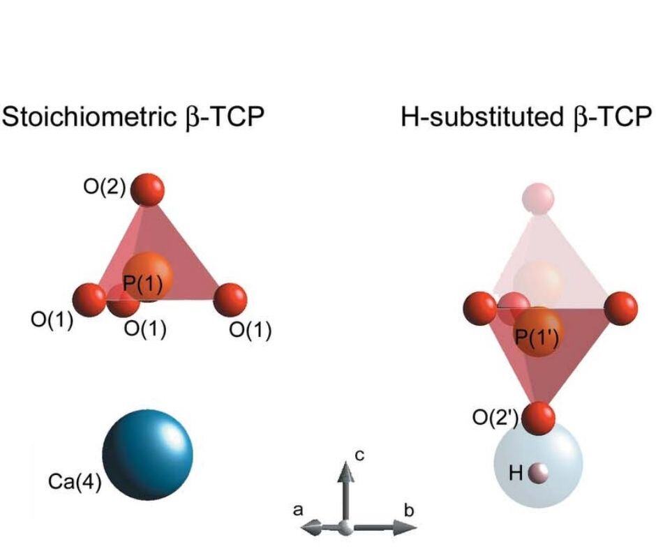
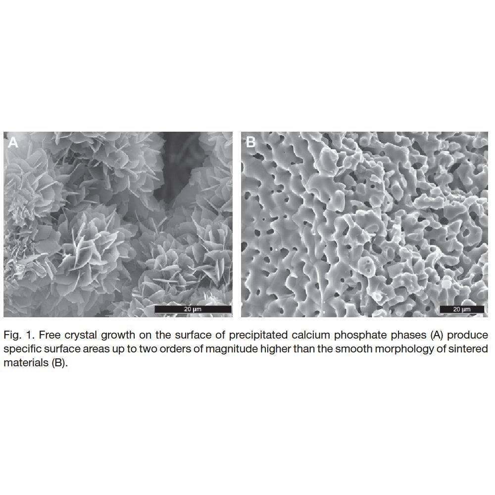
β-tricalcium phosphate (β-TCP) is one the most used and potent synthetic bone graft substitute. It is not only osteoconductive, but also osteoinductive. These properties, combined with its cell-mediated resorption, allow full bone defects regeneration. Its clinical outcome is sometimes considered to be “unpredictable”, possibly due to a poor understanding of β-TCP physico-chemical properties: β-TCP crystallographic structure is not fully uncovered; recent results suggest that sintered β-TCP is coated with a Ca-rich alkaline phase; β-TCP apatite-forming ability and osteoinductivity may be enhanced by a hydrothermal treatment; β-TCP grain size and porosity are strongly modified by the presence of minute amounts of β-calcium pyrophosphate or hydroxyapatite impurities. The aim of the present article is to provide a critical, but still rather comprehensive review of the current state of knowledge on β-TCP, with a strong focus on its synthesis and physico-chemical properties, and their link to the in vivo response.
Statement of significance
The present review documents the richness, breadth, and interest of the research devoted to β-tricalcium phosphate (β-TCP). β-TCP is synthetic, osteoconductive, osteoinductive, and its resorption is cell-mediated, thus making it one of the most potent bone graft substitutes. This comprehensive review reveals that there are a number of aspects, such as surface chemistry, crystallography, or stoichiometry deviations, that are still poorly understood. As such, β-TCP is still an exciting scientific playground despite a 50 year long history and > 200 yearly publications.
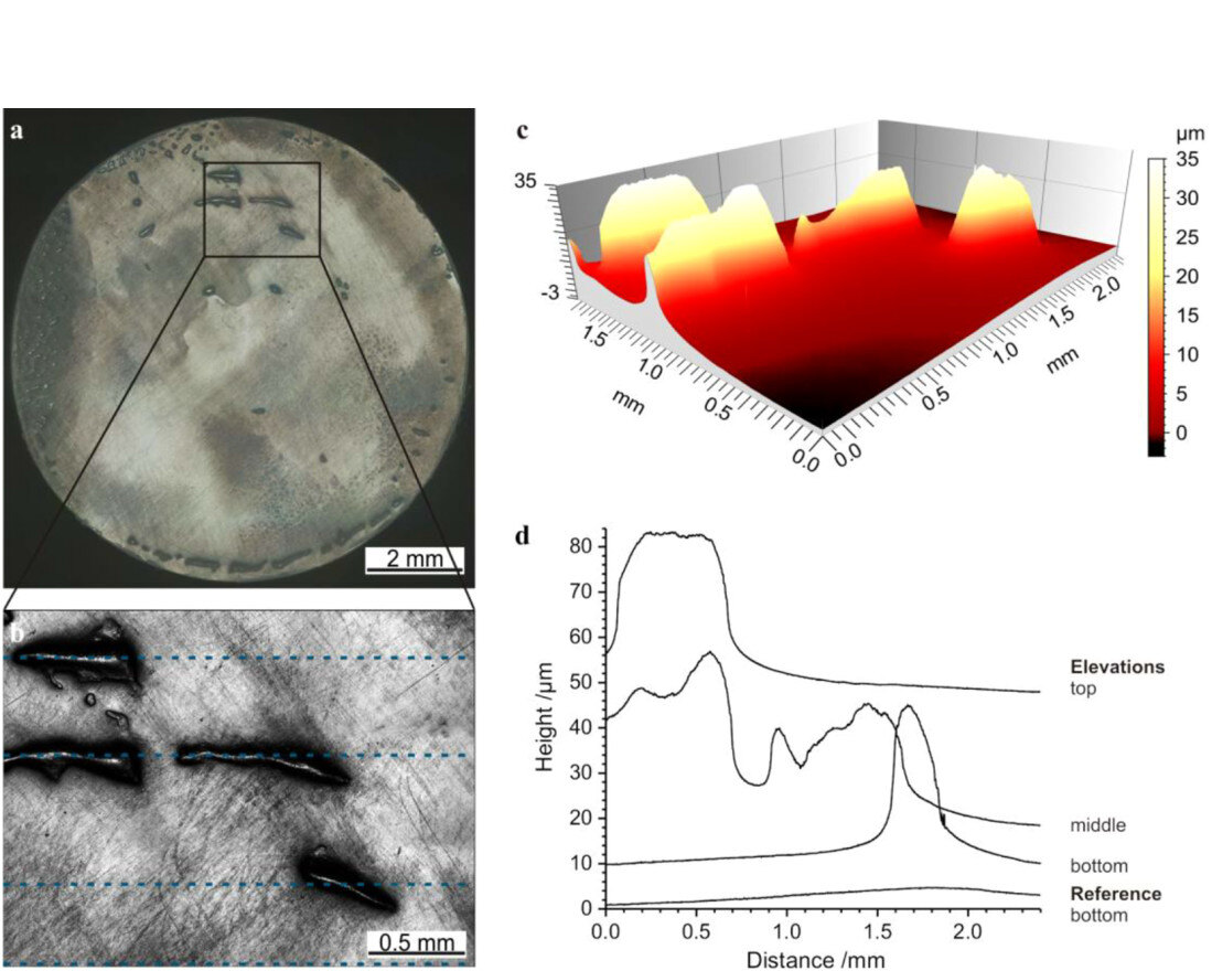
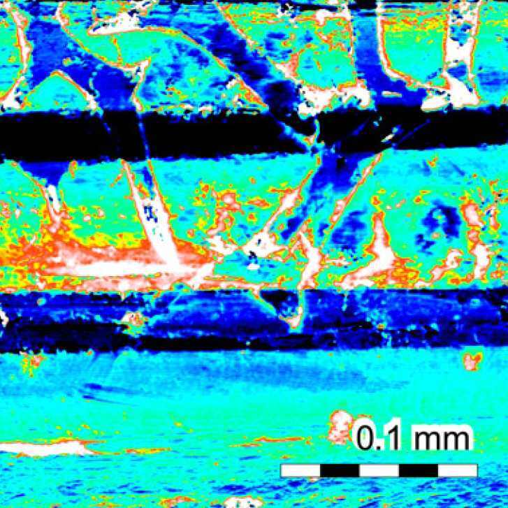
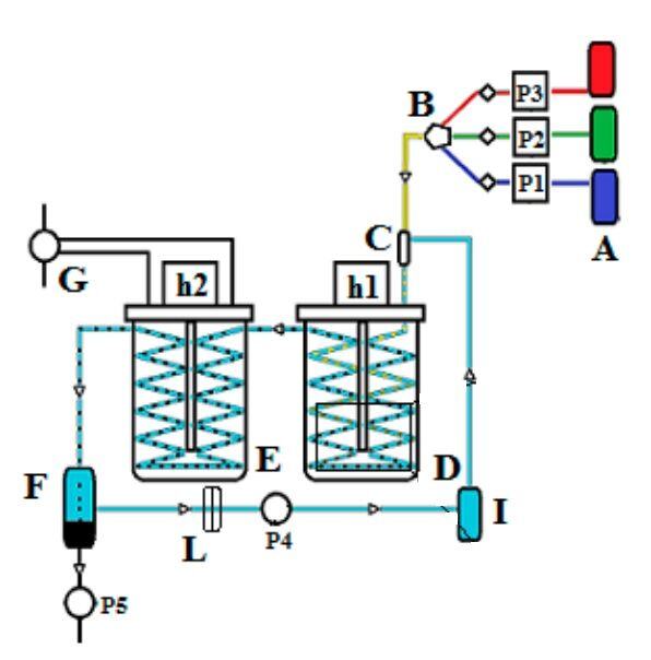
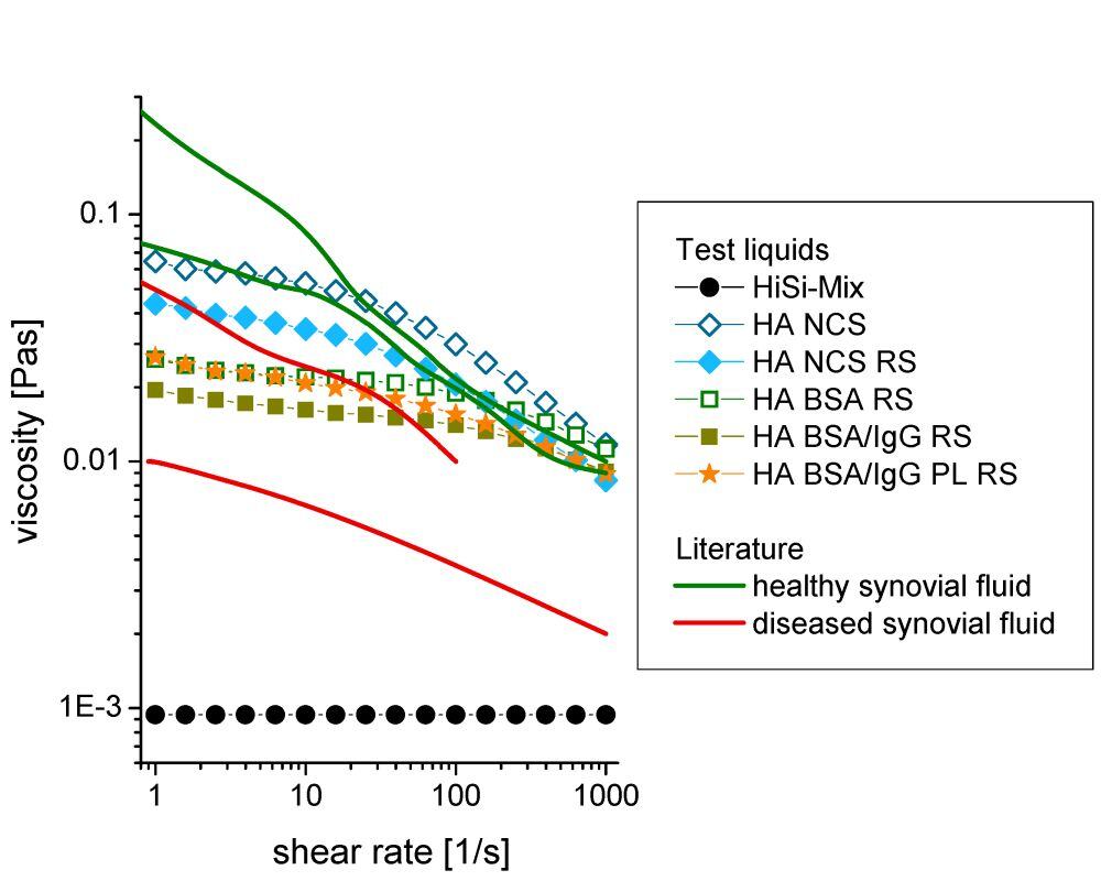
Protuberances on the surface of ultra-high-molecular-weight polyethylene (UHMWPE) pins were chemically and mechanically investigated in order to better understand the tribology of UHMWPE vs. CoCrMo, which is a typical material pairing for joint replacements.
Pin-on-disc wear tests were performed using pins made of UHMWPE articulating against discs made of a CoCrMo alloy. Wear tests were performed using two different test fluids: a standard test liquid used for hip-simulator tests and a synthetic synovial fluid containing hyaluronic acid, albumin, immunoglobulin G, the phospholipid lecithin and additionally sodium azide to fight bacterial growth.
After the wear tests, the pin surfaces exhibited scratches as well as protuberances with a pitting-like appearance. These protuberances, i.e. elevations protruding from the articulating surface, were 6 ± 3 μm high on the pins lubricated with the standard test liquid and 20 ± 5 μm high under the lubrication with the synthetic synovial fluid. Investigating the protuberances using Fourier transform infrared spectroscopy (FTIR) and scanning electron microscopy (SEM) showed that these were composed mainly of UHMWPE, together with amine groups from proteins. To our knowledge, the mechanical properties and namely shear resistance of these protuberances were investigated the first time. The hardness and the elastic modulus of the protuberances were similar to the bulk material, as revealed by nanoindentation. The shear resistance of the protuberances as measured by a nanoscratch test method was comparable or even higher than that of the bulk material.
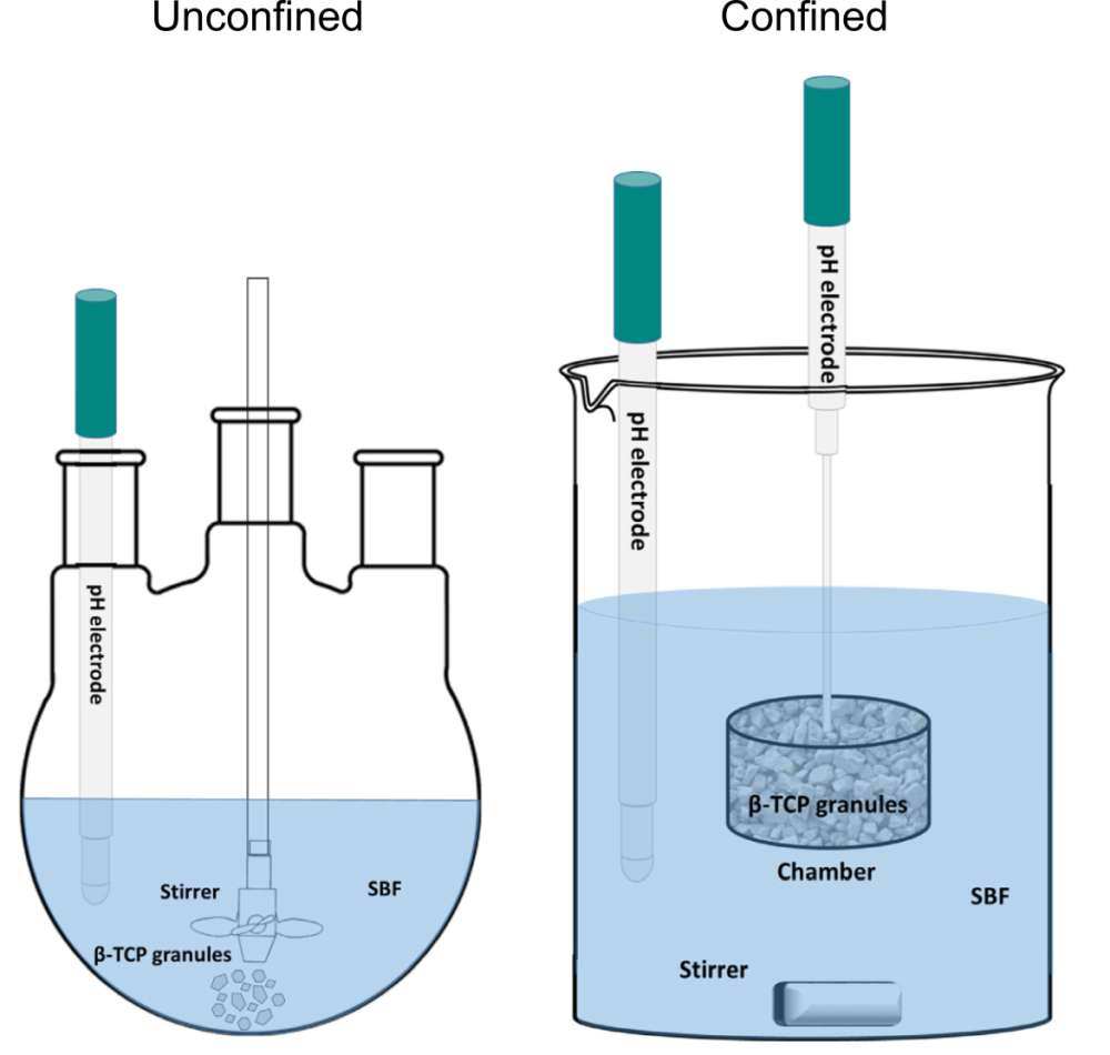
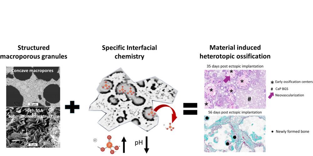
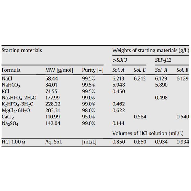
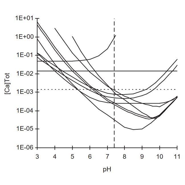
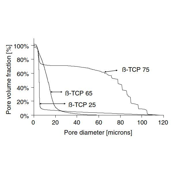
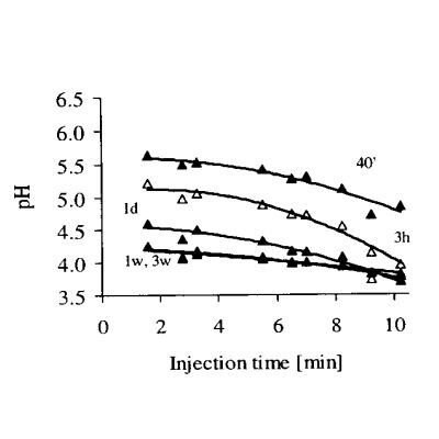
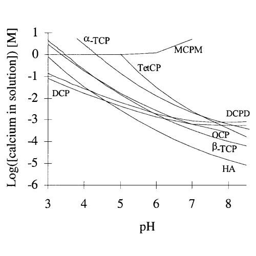
Several mechanisms proposed to explain the osteoinductive potential of calcium phosphates involve surface mineralization (“bioactivity”) and mention the occurrence of concentration gradients between the inner and the outer part of the implanted material. Determining the evolution of the local chemical environment occurring inside the pores of an implanted bone graft substitute (BGS) is therefore highly relevant. A quantitative and fast method was developed to measure the chemical changes occurring within the pores of β-Tricalcium Phosphate (β-TCP) granules incubated in a simulated body fluid. A factorial design of experiment was used to test the effect of particle size, specific surface area, microporosity, and purity of the β-TCP granules. Large pH, calcium and phosphate concentration changes were observed inside the BGS and lasted for several days. The kinetics and magnitude of these changes (up to 2 pH units) largely depended on the processing and properties of the granules. Interestingly, processing parameters that increased the kinetics and magnitude of the local chemical changes are parameters considered to favor calcium phosphate osteoinduction, suggesting that the model might be useful to predict the osteoinductive potential of BGSs.
Statement of significanceRecent results suggest that in situ mineralization of biomaterials (polymers, ceramics, metals) might be key in their ability to trigger ectopic bone formation. This is the reason why the effect on in situ mineralization of various synthesis parameters of β-tricalcium phosphate granules was studied (size, microporosity, specific surface area, and Ca/P molar ratio). To the best of our knowledge, this is the first article devoted to the chemical changes occurring within the pores of a bone graft substitute. We believe that the manuscript will prove to be highly important in the design and mechanistic understanding of drug-free osteoinductive biomaterials.
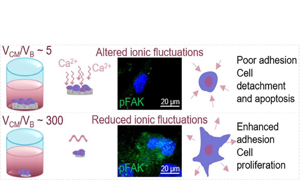
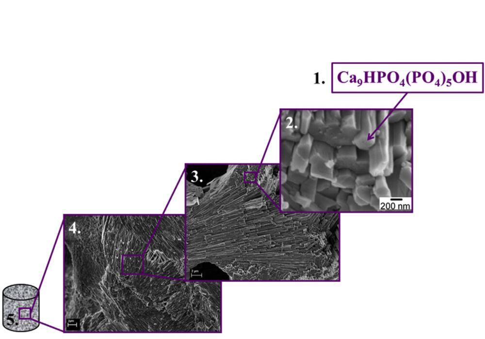
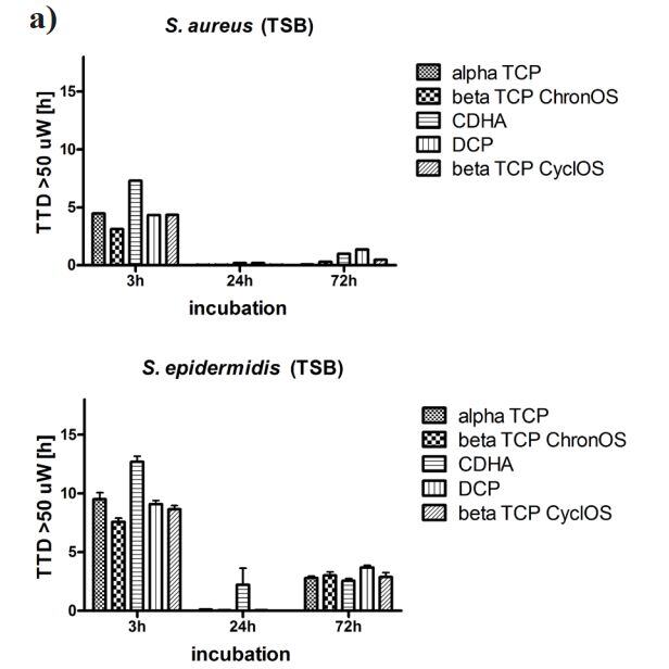
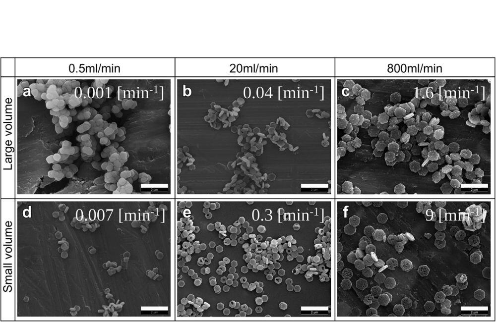
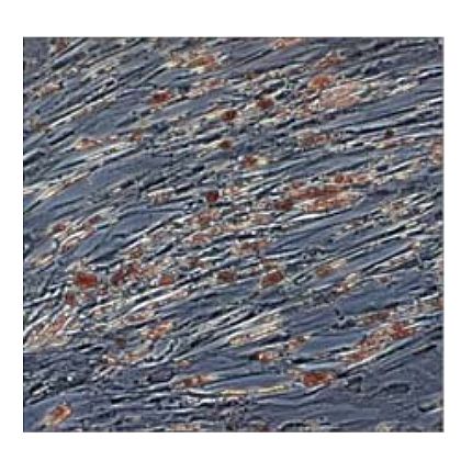
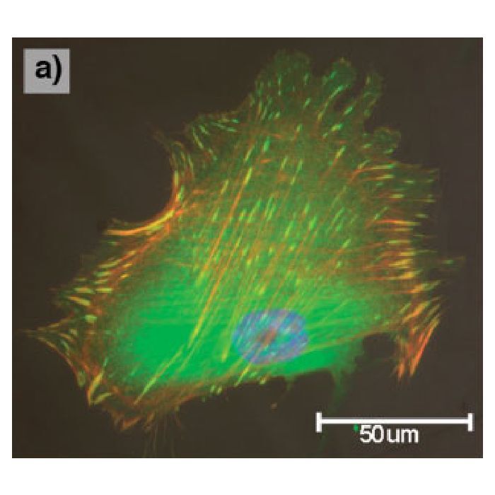
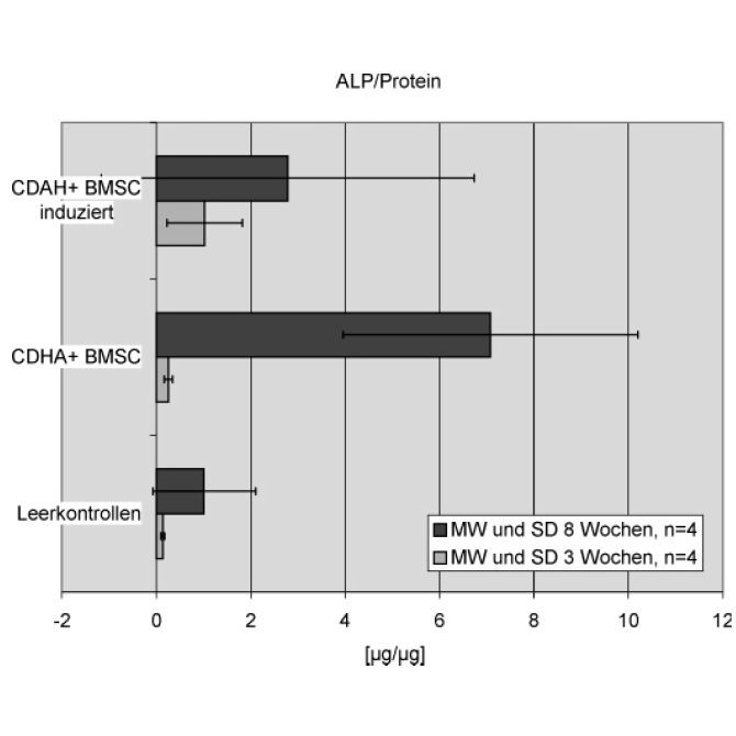
Biomaterials can interact with cells directly, that is, by direct contact of the cells with the material surface, or indirectly, through soluble species that can be released to or uptaken from the surrounding fluids. However, it is difficult to characterise the relevance of this fluid-mediated interaction separately from the topography and composition of the substrate, because they are coupled variables. These fluid-mediated interactions are amplified in the case of highly reactive calcium phosphates (CaPs) such as biomimetic calcium deficient hydroxyapatite (CDHA), particularly in static in vitro cultures. The present work proposes a strategy to decouple the effect of ion exchange from topographical features by adjusting the volume ratio between the cell culture medium and biomaterial (VCM/VB). Increasing this ratio allowed mitigating the drastic ionic exchanges associated to the compositional changes experienced by the material exposed to the cell culture medium. This strategy was validated using rat mesenchymal stem cells (rMSCs) cultured on CDHA and beta-tricalcium phosphate (β-TCP) discs using different VCM/VB ratios. Whereas in the case of β-TCP the cell response was not affected by this ratio, a significant effect on cell adhesion and proliferation was found for the more reactive CDHA. The ionic exchange, produced by CDHA at low VCM/VB, altered cell adhesion due to the reduced number of focal adhesions, caused cell shrinkage and further rMCSs apoptosis. This was mitigated when using a high VCM/VB, which attenuated the changes of calcium and phosphate concentrations in the cell culture medium, resulting in rMSCs spreading and a viability over time. Moreover, rMSCs showed an earlier expression of osteogenic genes on CDHA compared to sintered β-TCP when extracellular calcium fluctuations were reduced.
Fluid mediated interactions play a significant role in the bioactivity of calcium phosphates. Ionic exchange is amplified in the case of biomimetic hydroxyapatite, which makes the in vitro characterisation of cell-material interactions especially challenging. The present work proposes a novel and simple strategy to explore the mechanisms of interaction of biomimetic and sintered calcium phosphates with mesenchymal stem cells. The effects of topography and ion exchange are analysed separately by modifying the volume ratio between cell culture medium and biomaterial. High ionic fluctuations interfered in the maturation of focal adhesions, hampering cell adhesion and leading to increased apoptosis and reduced proliferation rate.
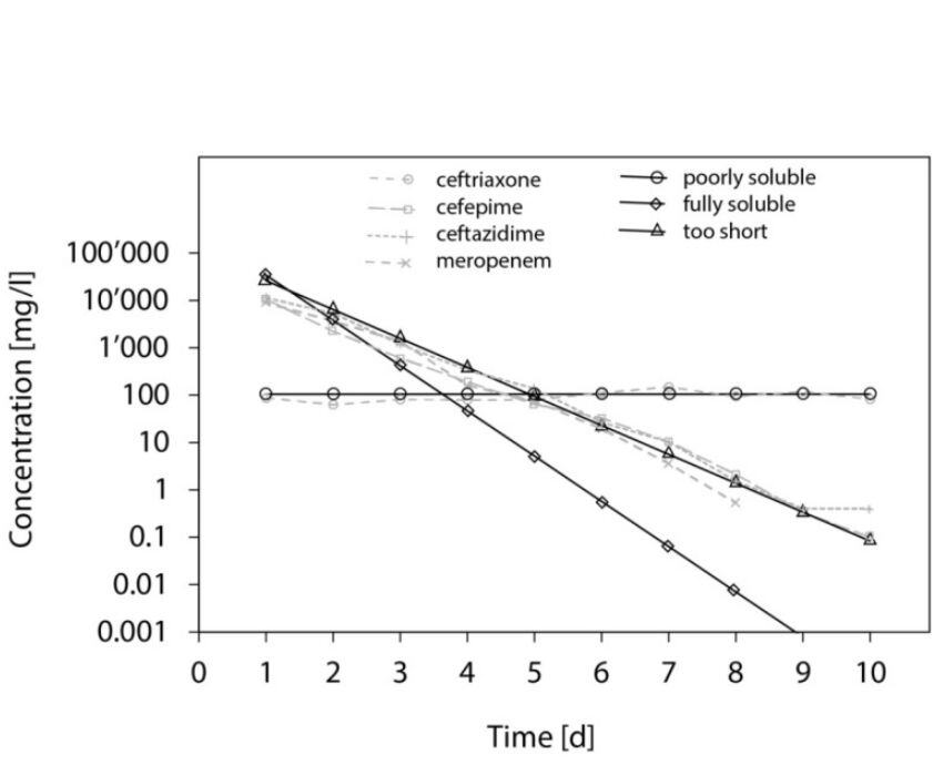
Introduction: Local application of antibiotics provides high concentrations at the site of interest, with minimal systemic toxicity. Carrier materials might help manage dead space. Calcium sulphate (CaSO4) has a dissolution time that only slightly exceeds the usually recommended duration of systemic antibiotic treatments. This in vitro study evaluates compatibility, release kinetics and antibacterial activity of new combinations of antibiotics with CaSO4 as carrier material.
Methods: CaSO4 pellets added with 8% w/w antibiotic powder were exposed once in phosphate-buffered saline (PBS) solution and once in bovine plasma, in an elution experiment run over 6 weeks at 37 °C. Antibiotic elution was examined at various time points. Concentration was measured by liquid chromatography with tandem mass spectrometry. Antimicrobial activity was checked with an agar diffusion test.
Results: Piperacillin-tazobactam, ceftazidime, cefepime, and meropenem showed fast reduction of concentration and activity. Flucloxacillin and cefuroxime remained present in relevant concentrations for 4 weeks. Ciprofloxacin, levofloxacin and clindamycin lasted for 6 weeks, but also at cell toxic concentrations. Ceftriaxone showed a near-constant release with only a small reduction of concentration from 130 to 75 mg/l. Elution profiles from PBS and plasma were comparable.
Conclusion: CaSO4 provides new possibilities in the local treatment of bone and joint infections. Ceftriaxone appears to be of particular interest in combination with CaSO4. Release persists at clinically promising concentrations, and appears to have a depot-like slow release from CaSO4, with only a small reduction in activity and concentration over 6 weeks. To the best of our knowledge, such a particular persistent release never was described before, for any antibiotic in combination with a carrier material for local application.
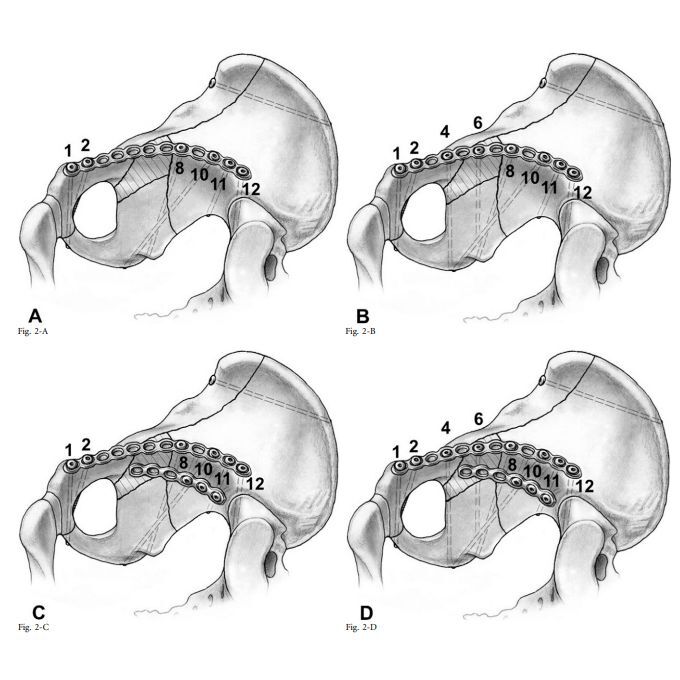
Background: In elderly patients who have sustained an acetabular fracture involving disruption of the quadrilateral plate (QLP), postoperative loading of the joint beyond the level of partial weight-bearing can result in medial redisplacement of the QLP. The purpose of this biomechanical study was to compare the performances of 4 different fixation constructs intended to prevent medial redisplacement of the QLP.
Methods: Anterior column posterior hemitransverse (ACPHT) fractures with disruption of the QLP were created on synthetic hemipelves (fourth-generation Sawbones models) and subsequently stabilized with (1) a 12-hole plate bridging the QLP (Group 1), (2) the plate with added periarticular screws along the QLP (Group 2), (3) the plate combined with an infrapectineal buttress plate (Group 3), or (4) the plate with the added periarticular screws as well as the buttress plate (Group 4). The point of load application on the acetabulum was defined to be the same as the point of application of maximum vertical hip contact force during normal walking. Loads were applied to simulate either partial weight-bearing (20 cycles, from 35 to 350 N) or inadvertent supraphysiologic loads (linearly increasing loads until the onset of failure, defined as fragment displacement of >3 mm). A universal testing machine was synchronized with a digital image correlation system to optically track redisplacement at the QLP. The level of significance was set at p < 0.05.
Results: During experimental simulation of partial weight-bearing, maximum fracture step openings never exceeded 2 mm. During simulation of inadvertent supraphysiologic load, the median load to failure was higher (p < 0.05) in Group 2 (962 N; range, 798 to 1,000 N) and Group 4 (985 N; range, 887 to 1,000 N) compared with Group 1 (445 N; range, 377 to 583 N) and Group 3 (671 N; range, 447 to 720 N).
Conclusions: All 4 fixation constructs performed in an acceptable manner on testing with simulated partial weight-bearing. Only additional periarticular screws along the QLP increased the fixation strength.
Clinical Relevance: Redisplacement of the QLP resulting in an incongruency of the hip joint has been associated with poor long-term outcomes. Within the constraints of this study, periarticular long screws were superior to infrapectineal buttress plates in preventing medial redisplacement of the QLP.
Objective
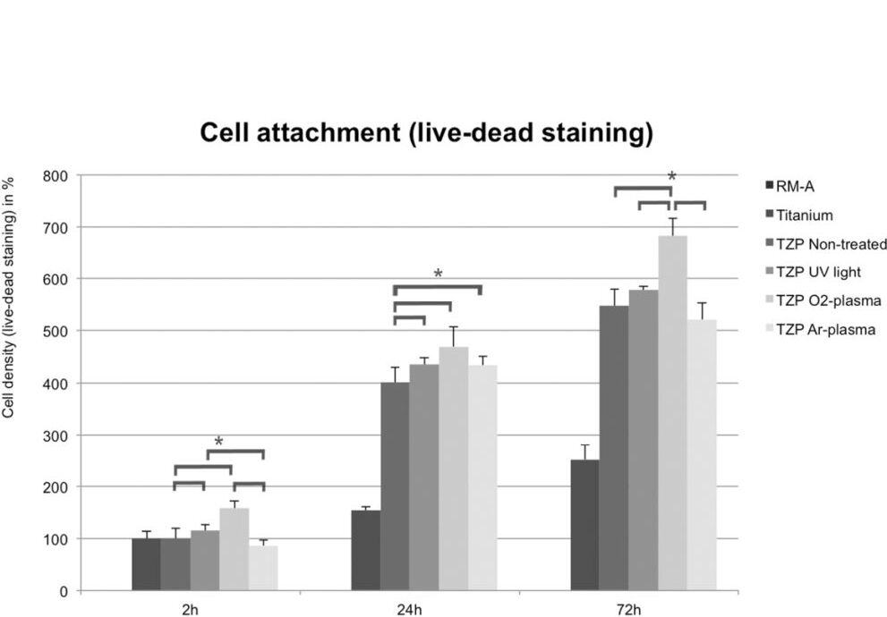
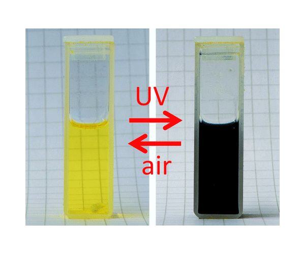
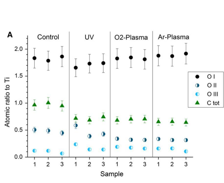
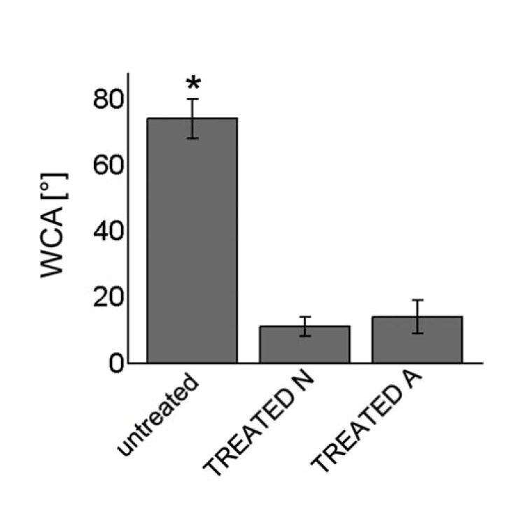
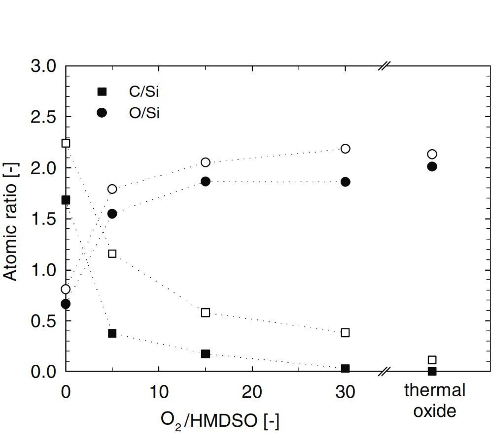
The aim of this study was to compare UV light and non-thermal plasma (NTP) treatment regarding the improvement of physical material characteristics and cell reaction on titanium surfaces in vitro after short-term functionalization.
Materials and methods
Moderately rough (Ra 1.8–2.0 μm) sandblasted and acid-etched titanium disks were treated by UV light (0.05 mW/cm2 at λ = 360 nm and 2 mW/cm2 at λ = 250 nm) or by NTP (24 W, -0.5 mbar) of argon or oxygen for 12 min each. Surface structure was investigated by scanning electron microscopy, confocal microscopy and X-ray photoelectron spectroscopy (XPS). Hydrophilicity was assessed by dynamic contact angle measurement. Cell attachment, viability, cell proliferation and cytotoxicity were assessed in vitro using murine osteoblast-like cells.
Results
UV irradiation or NTP treatment of titanium surfaces did not alter the surface structure. XPS analysis revealed a significantly increased oxidation of the surface and a decrease of carbon after the use of either method. NTP and UV light led to a significant better cell attachment of murine osteoblasts; significantly more osteoblasts grew on the treated surfaces at each time point (p < 0.001).
Conclusions
UV light as well as NTP modified the surface of titanium and significantly improved the conditions for murine osteoblast cells in vitro. However, results indicate a slight advantage for NTP of argon and oxygen in a short time interval of surface functionalization compared to UV.
Clinical relevance
UV light and NTP are able to improve surface conditions of dental implants made of titanium.
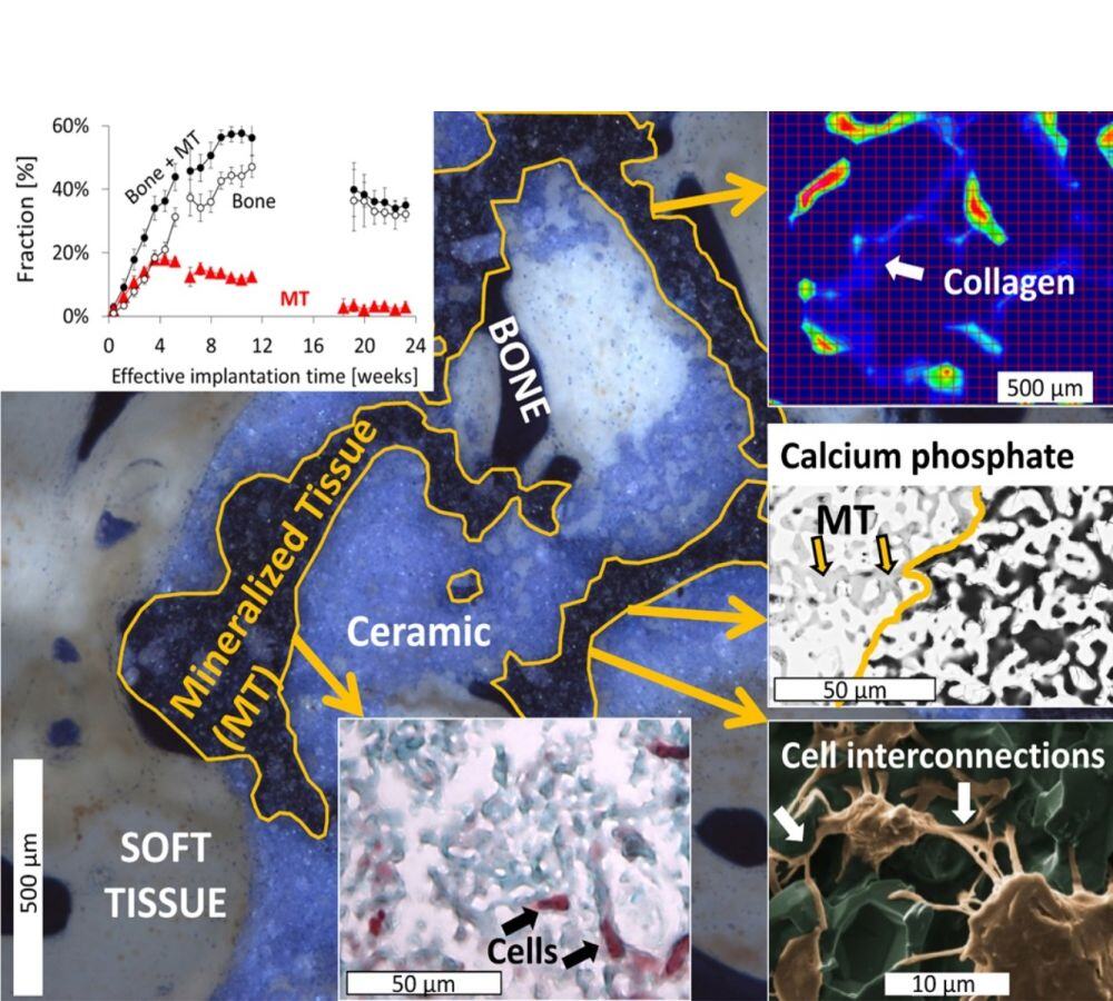
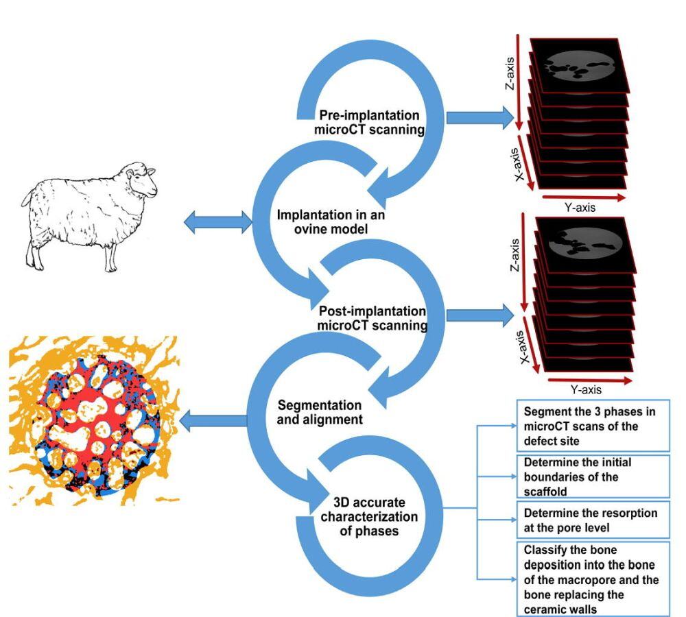
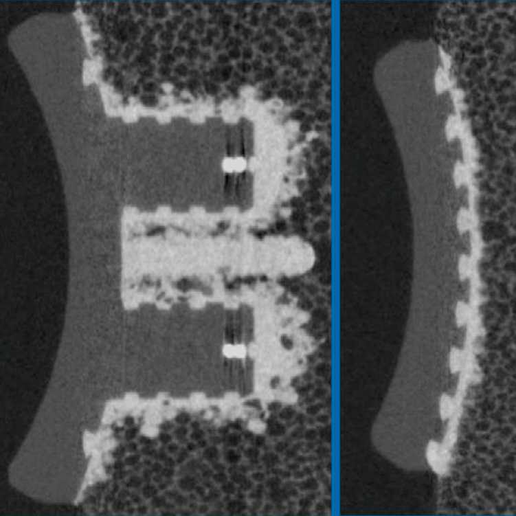
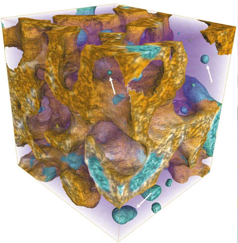
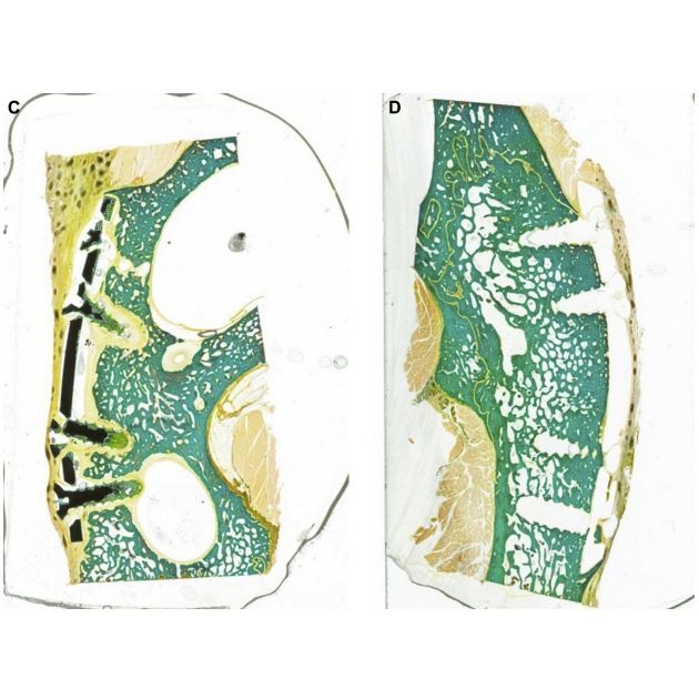
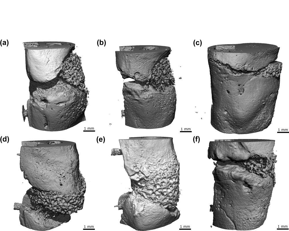
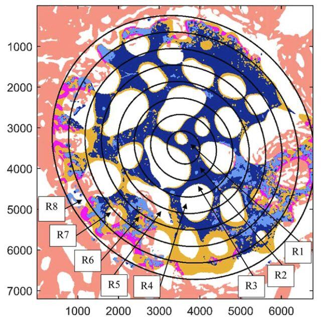
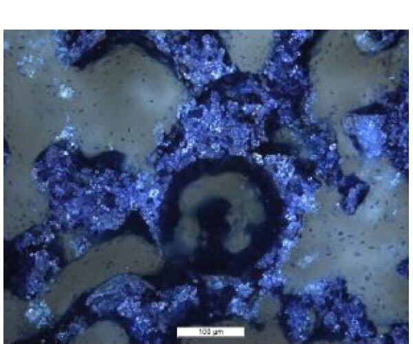
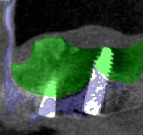
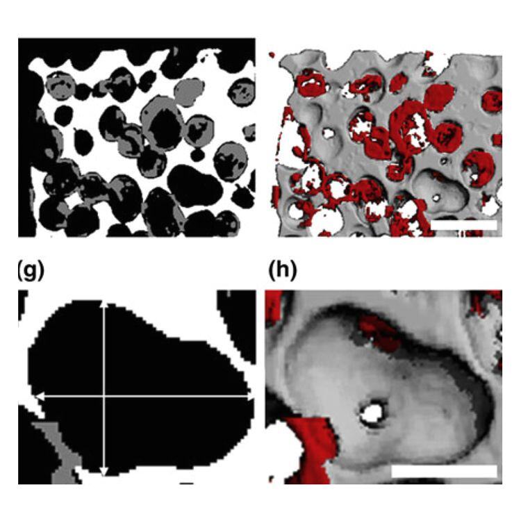
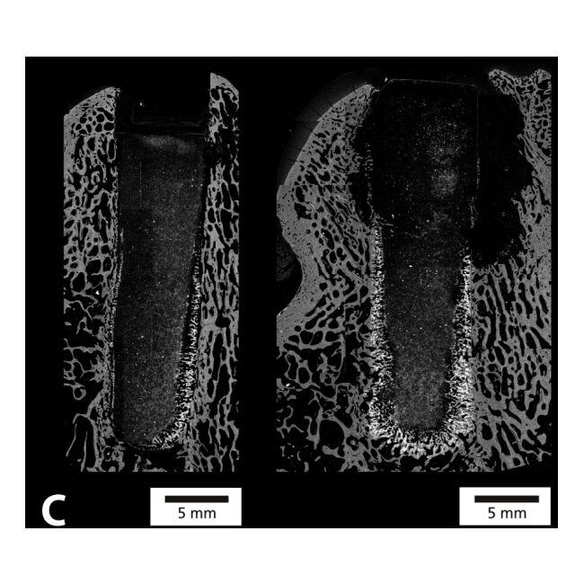
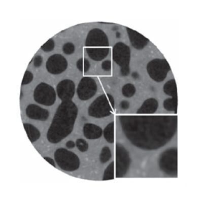
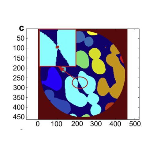
Micro-computed tomography (microCT) is commonly used to characterize the three-dimensional structure of bone graft scaffolds before and after implantation in order to assess changes occurring during implantation. The accurate processing of the microCT datasets of explanted β-tricalcium phosphate (β-TCP) scaffolds poses significant challenges because of (a) the overlap in the grey values distribution of ceramic remnants, bone, and soft tissue, and of (b) the resorption of the bone substitute during the implantation. To address those challenges, this article introduces and rigorously validates a new processing technique to accurately distinguish these three phases found in the explanted β-TCP scaffolds. Specifically, the microCT datasets obtained before and after implantation of β-TCP scaffolds were aligned in 3D, and the characteristic grey value distributions of the three phases were extracted, thus allowing for (i) the accurate differentiation between these three phases (ceramic remnants, bone, soft tissue), and additionally for (ii) the localization of the defect site in the post-implantation microCT dataset. Using the similarity matrix, a 94±1% agreement was found between algorithmic results and the visual assessment of 556,800 pixels. Moreover, the comparison of the segmentation results of the same microCT and histology section further confirmed the validity of the present segmentation algorithm. This new technique could lead to a more common use of microCT in analyzing the complex 3D processes and to a better understanding of the biological processes occurring after the implantation of ceramic bone graft substitutes.
STATEMENT OF SIGNIFICANCE:
Calcium-phosphate scaffolds are being increasingly used to repair critical bone defects. Methods for the accurate characterization of the repair process are still lacking. The present study introduced and validated a novel image-processing technique, using micro-computed tomography (mCT) datasets, to investigate material phases present in biopsies. Specifically, the new method combined mCT datasets from the scaffold before and after implantation to access the characteristic data of the ceramic for more accurate analysis of bone biopsies, and as such to better understand the interactions of the scaffold design and the bone repair process.
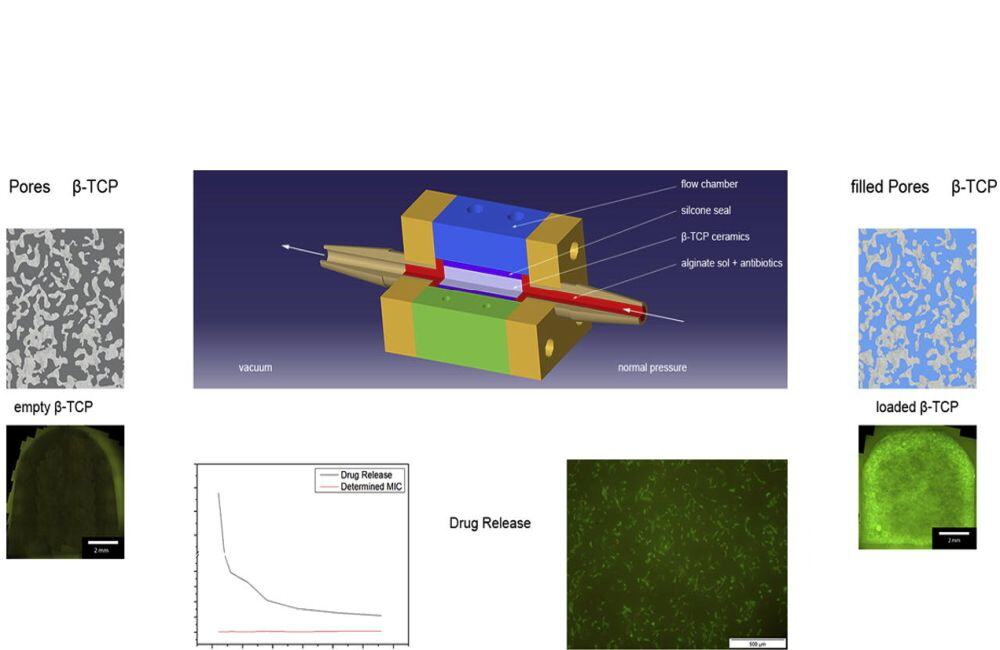
Objective
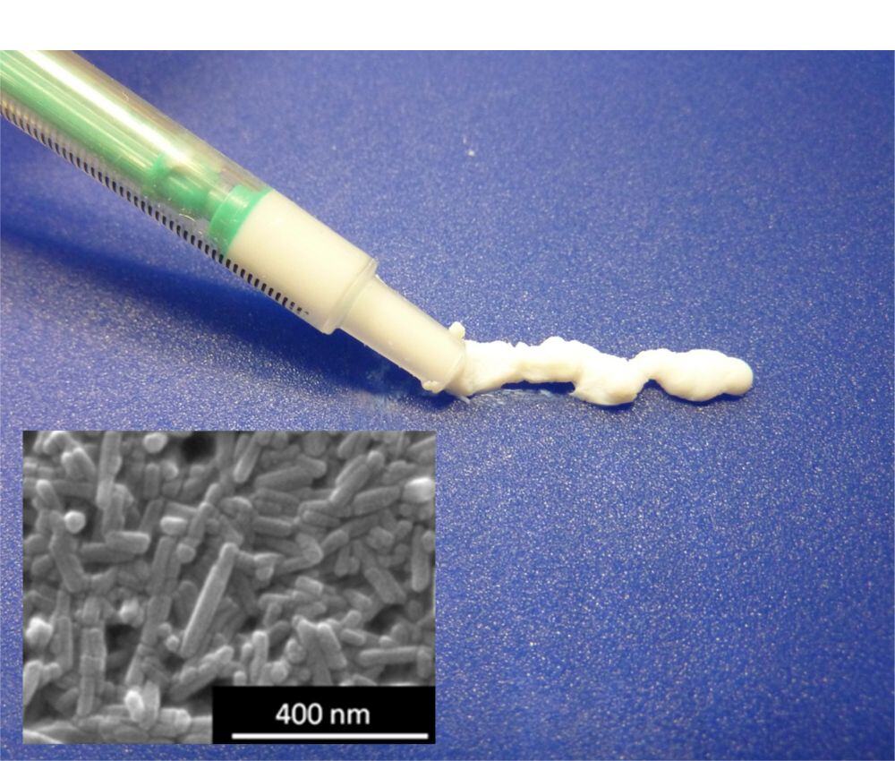
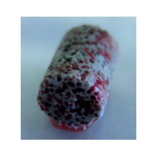
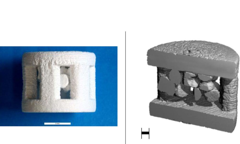
The aim of this study was to produce a novel composite of microporous β-TCP filled with alginate and Vancomycin (VAN) to prolong the release behavior of the antibiotic for up to 28 days.
Material and methods
Using the flow chamber developed by the group, porous ceramics in a directional flow were filled with alginates of different composition containing 50 mg/mL of antibiotics. After cross-linking the alginate with calcium ions, incubation took place in 10 mL double-distilled water for 4 weeks at 37 °C. At defined times (1, 2, 3, 6, 9, 14, 20 and 28 days), the liquid was completely exchanged and analyzed by capillary zone electrophoresis and microtiter trials. For statistical purposes, the mean and standard deviation were calculated and analyzed by ANOVA.
Results
The release of VAN from alginate was carried out via an external calcium source over the entire period with concentrations above the minimal inhibitory concentration (MIC). The burst release measured 35.2 ± 1.5%. The release of VAN from alginate with an internal calcium source could only be observed over 14 days. The burst release here was 61.9 ± 4.3%. The native alginate’s burst release was 54.1 ± 7.8%; that of the sterile alginate 40.5 ± 6.4%. The microtiter experiments revealed efficacy over the entire study period for VAN. The MIC value was determined in the release experiments as well in a range of 0.5–2.0 μg/mL against Staphylococcus aureus.
Statement of Significance
Drug release systems based on β-TCP and hydrogels are well documented in literature. However, in all described systems the ceramic, as granule or powder, is inserted into a hydrogel. In our work, we do the opposite, a hydrogel which acts as reservoir for antibiotics is placed into a porous biodegradable ceramic. Eventually, this system should be applied as treatment of bone infections. Contrary to the “granule in hydrogel” composites it has the advantage of mechanical stability. Thus, it can take over functions of the bone during the healing process. For a quicker translation from our scientific research into clinical use, only FDA approved materials were used in this work.
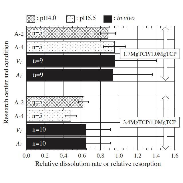
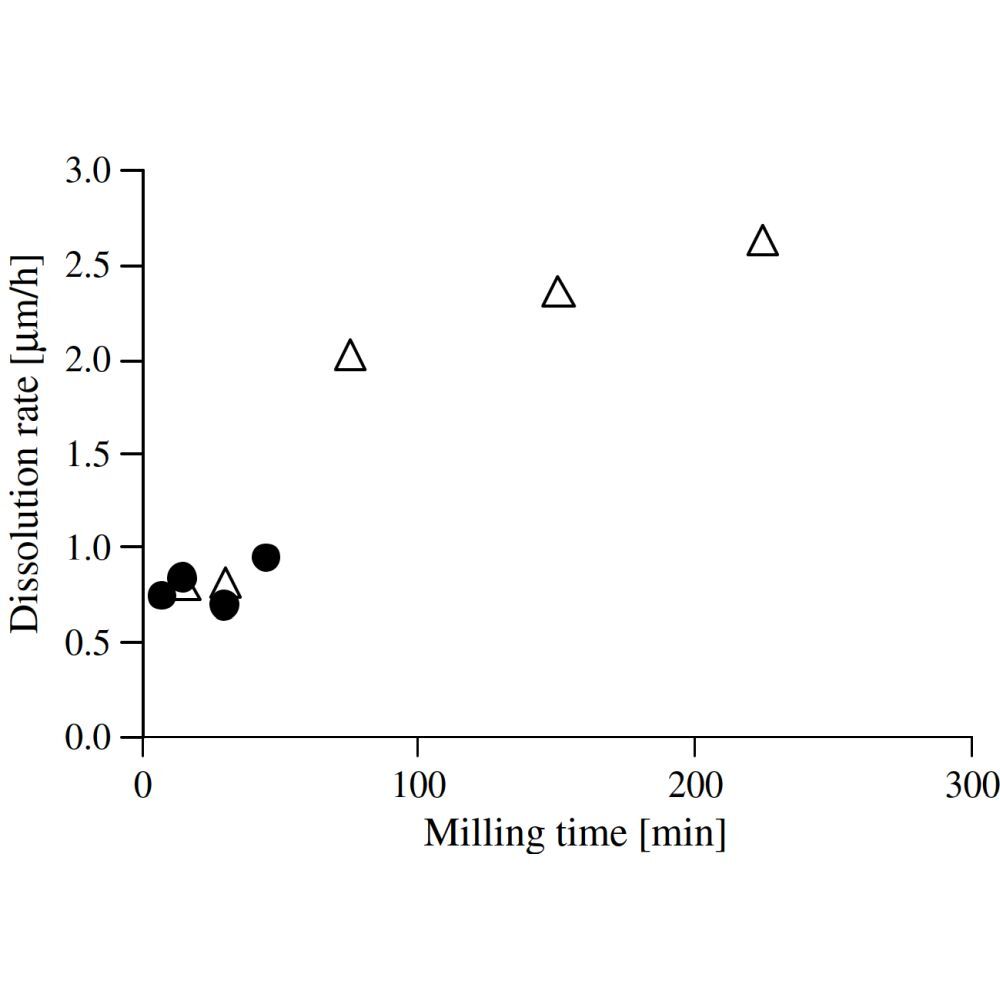
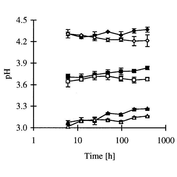
A potential standard method for measuring the relative dissolution rate to estimate the resorbability of calcium-phosphate-based ceramics is proposed. Tricalcium phosphate (TCP), magnesium-substituted TCP (MgTCP) and zinc-substituted TCP (ZnTCP) were dissolved in a buffer solution free of calcium and phosphate ions at pH 4.0, 5.5 or 7.3 at nine research centers. Relative values of the initial dissolution rate (relative dissolution rates) were in good agreement among the centers. The relative dissolution rate coincided with the relative volume of resorption pits of ZnTCP in vitro. The relative dissolution rate coincided with the relative resorbed volume in vivo in the case of comparison between microporous MgTCPs with different Mg contents and similar porosity. However, the relative dissolution rate was in poor agreement with the relative resorbed volume in vivo in the case of comparison between microporous TCP and MgTCP due to the superimposition of the Mg-mediated decrease in TCP solubility on the Mg-mediated increase in the amount of resorption. An unambiguous conclusion could not be made as to whether the relative dissolution rate is predictive of the relative resorbed volume in vivo in the case of comparison between TCPs with different porosity. The relative dissolution rate may be useful for predicting the relative amount of resorption for calcium-phosphate-based ceramics having different solubility under the condition that the differences in the materials compared have little impact on the resorption process such as the number and activity of resorbing cells.
Statement of significance
The evaluation and subsequent optimization of the resorbability of calcium phosphate are crucial in the use of resorbable calcium phosphates. Although the resorbability of calcium phosphates has usually been evaluated in vivo, establishment of a standard in vitro method that can predict in vivo resorption is beneficial for accelerating development and commercialization of new resorbable calcium phosphate materials as well as reducing use of animals. However, there are only a few studies to propose such an in vitro method within which direct comparison was carried out between in vitro and in vivo resorption. We propose here an in vitro method based on measuring dissolution rate. The efficacy and limitations of the method were evaluated by international round-robin tests as well as comparison with in vivo resorption studies for future standardization. This study was carried out as one of Versailles Projects on Advanced Materials and Standards (VAMAS).

Aim
To test in vitro the mechanical resistance, rotational misfit and failure mode of three original implant-abutment connections and to compare them to two connections between non-original abutments connected to one of the original implants.
Material and Methods
Three different implants with small diameters (3.3 mm for Straumann Roxolid, 3.5 mm for Nobel Biocare Replace and Astra Tech Osseospeed TX) were connected with individualized titanium abutments.
Twelve implants from each system were connected to their original abutments (Straumann CARES, Nobel Biocare Procera, Astra Tech Atlantis). Twenty-four Roxolid implants were connected with non-original abutments using CAD/CAM procedures from the other two manufacturers (12 Nobel Biocare Procera and 12 Astra Tech Atlantis). For the critical bending test, a Zwick/Roell 1475 machine and the Xpert Zwick/Roell software were used.
Results
The rotational misfit varied when comparing the different interfaces. The use of non-original grade V titanium abutments on Roxolid implants increased the force needed for deformation. The fracture mode was different with one of the original connections.
Conclusions
Non-original abutments differ in design of the connecting surfaces and material and demonstrate higher rotational misfit. These differences may result in unexpected failure modes.
Background
Finding the right balance between tibial coverage and minimal implant overhang is an important factor in TKA. Another significant cause of failure is component malrotation.
Methods
An average master shape of the proximal tibia at TKA resection level was calculated using fine slice computed tomographies of 117 cadaveric knees. To find out whether alternate implant contours would be necessary depending on the patient's body size, we established five subgroups to compare. CAD-Analysis was performed to simulate the overhang produced after ±4°/±7°/±10° rotation.
Results
A master shape for the tibial resection cut (with a 5° posterior slope, 7mm under lateral joint line) could be determined. Neither left vs. right knee joint, nor male vs. female nor the size subdivision appears to alter the calculated master shape significantly. The optimized shape allowing for ±4° of rotational freedom was found to be the best variant.
Conclusions
Valid methods have been obtained to design a two-dimensional average shape of the tibial plateau. The modifications described in this study might come in useful, when designing future implant designs.
Clinical relevance
An optimized fit at the tibial plateau and lower rates of component malrotation may result in better outcomes after TKA.
Purpose
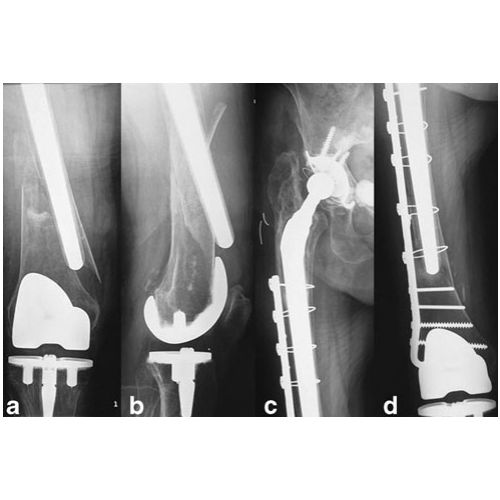
Fixation of periprosthetic hip fractures with intracortical anchorage might not be feasible in cases with bulky implants and/or poor bone stock.
Methods
Rotational stability of new plate inserts with extracortical anchorage for cerclage fixation was measured and compared to the stability found using a standard technique in a biomechanical setup using a torsion testing machine. In a synthetic PUR bone model, transverse fractures were fixed distally using screws and proximally by wire cerclages attached to the plates using “new” (extracortical anchorage) or “standard” (intracortical anchorage) plate inserts. Time to fracture consolidation and complications were assessed in a consecutive series of 18 patients (18 female; mean age 81 years, range 55–92) with periprosthetic hip fractures (ten type B1, eight type C-Vancouver) treated with the new device between July 2003 and July 2010.
Results
The “new” device showed a higher rotational stability than the “standard” technique (p < 0.001). Fractures showed radiographic consolidation after 14 ± 5 weeks (mean ± SD) postoperatively in patients. Revision surgery was necessary in four patients, unrelated to the new technique.
Conclusion
In periprosthetic hip fractures in which fixation with intracortical anchorage using conventional means might be difficult due to bulky revision stems and/or poor bone stock, the new device may be an addition to the range of existing implants.

Background and purpose
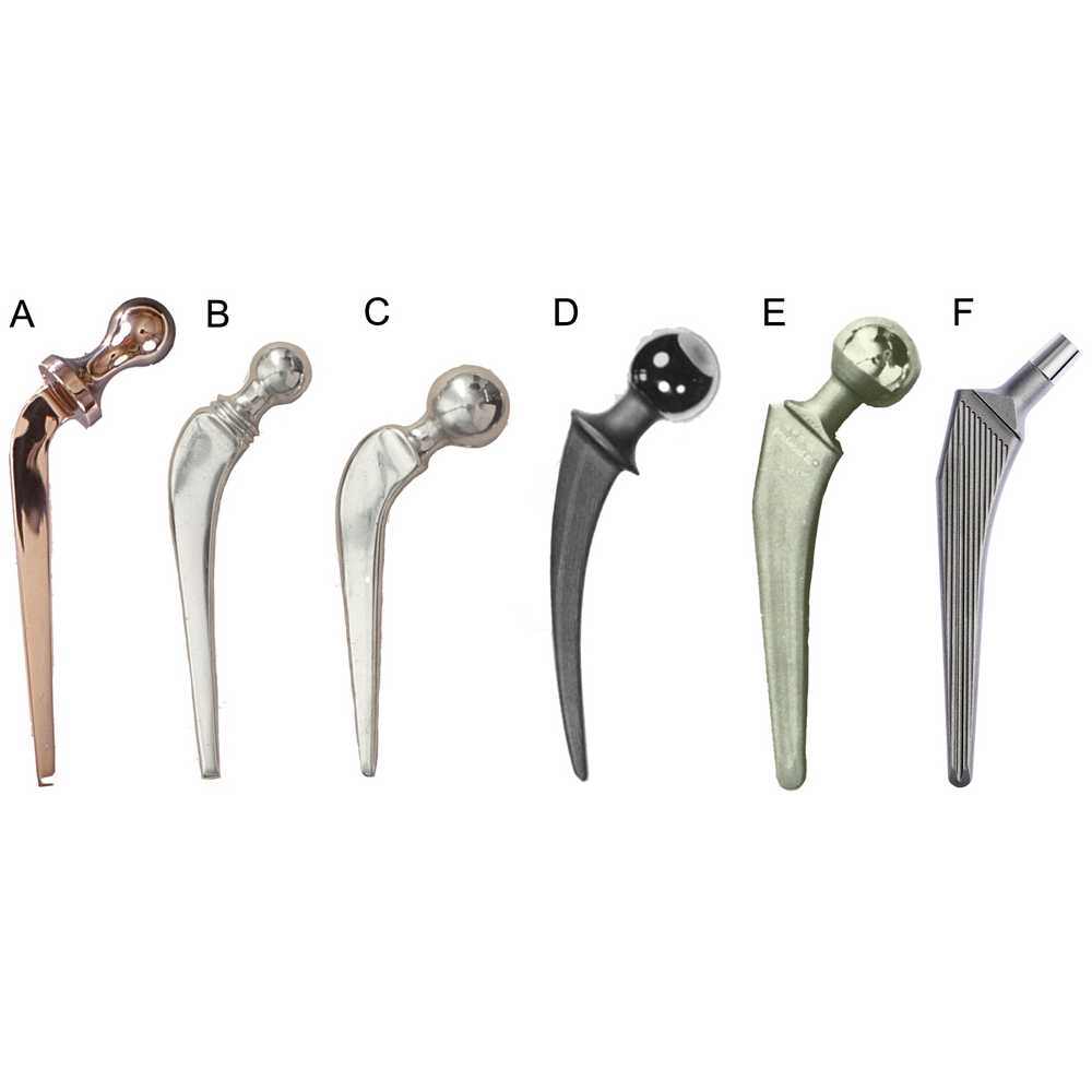
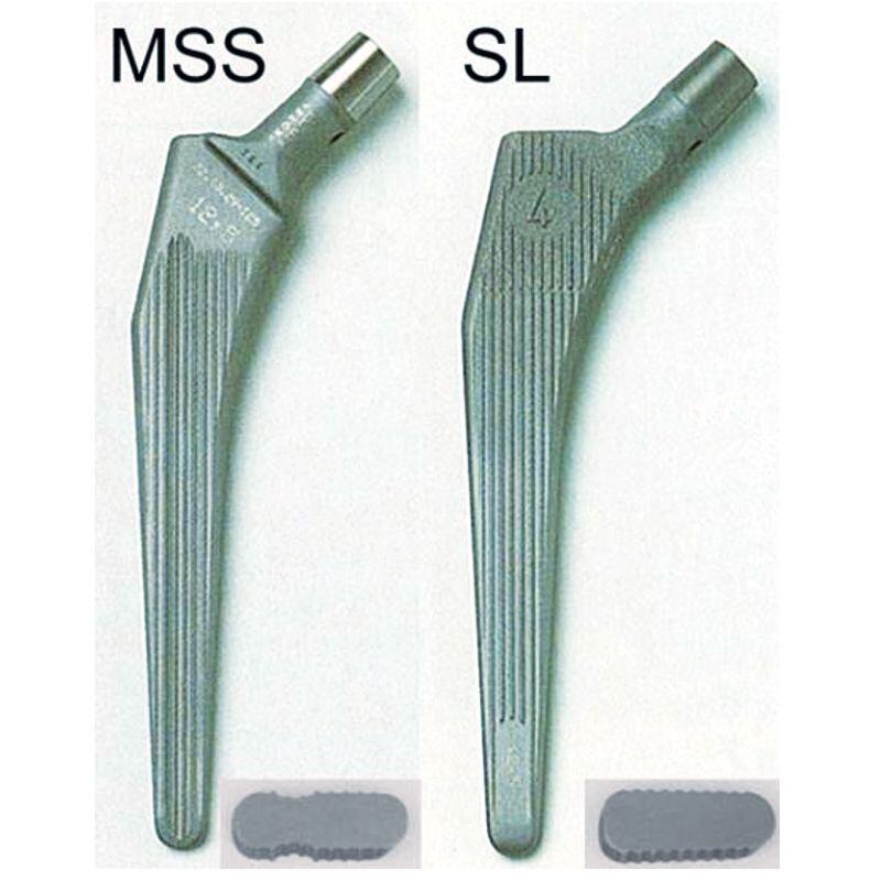
Even small differences in design variables for the femoral stem may influence the outcome of a hip arthroplasty. We performed a risk factor analysis for aseptic loosening of 4 different versions of cemented Müller-type straight stems with special emphasis on design modifications (2 shapes, MSS or SL, and 2 materials, CoNiCrMo (Co) or Ti-6Al-7Nb (Ti)).
Methods
We investigated 828 total hip replacements, which were followed prospectively in our in-house register. All stems were operated in the same setup, using Sulfix-6 bone cement and a second-generation cementing technique. Demographic and design-specific risk factors were analyzed using an adjusted Cox regression model.
Results
The 4 versions showed marked differences in 15-year stem survival with aseptic loosening as the endpoint: 94% (95% CI: 89–99) for MSS Co, 83% (CI: 75–91) for SL Co, 81% (CI: 76–87) for MSS Ti and 63% (CI: 56–71) for SL Ti. Cox regression analysis showed a relative risk (RR) for aseptic loosening of 3 (CI: 2–5) for stems made of Ti and of 2 (CI: 1–2) for the SL design. The RR for aseptic stem loosening increased to 8 (CI: 4–15) when comparing the most and the least successful designs (MSS Co and SL Ti).
Interpretation
Cemented Müller-type straight stems should be MSS-shaped and made of a material with high flexural strength (e.g. cobalt-chrome). The surface finish should be polished (Ra < 0.4 μm). These technical aspects combined with modern cement-ing techniques would improve the survival of Müller-type straight stems. This may be true for all types of cemented stems.


No Abstract Available

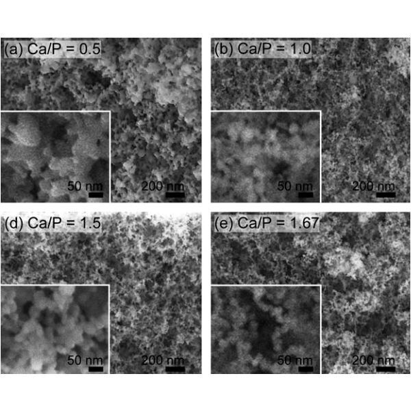
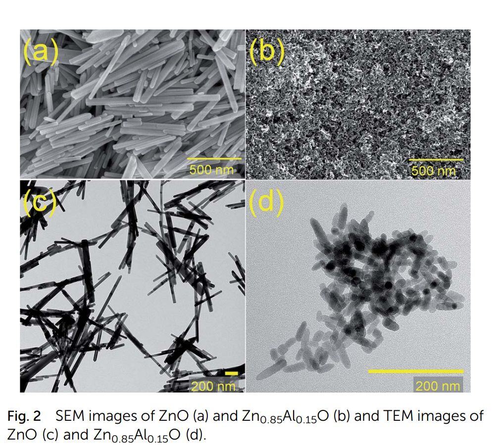
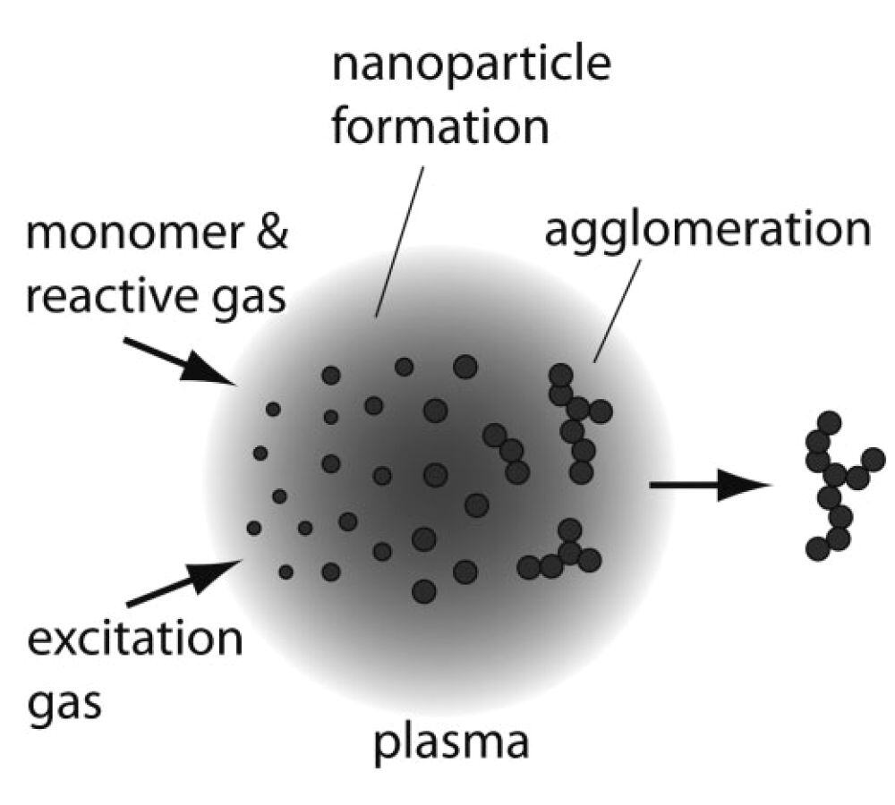
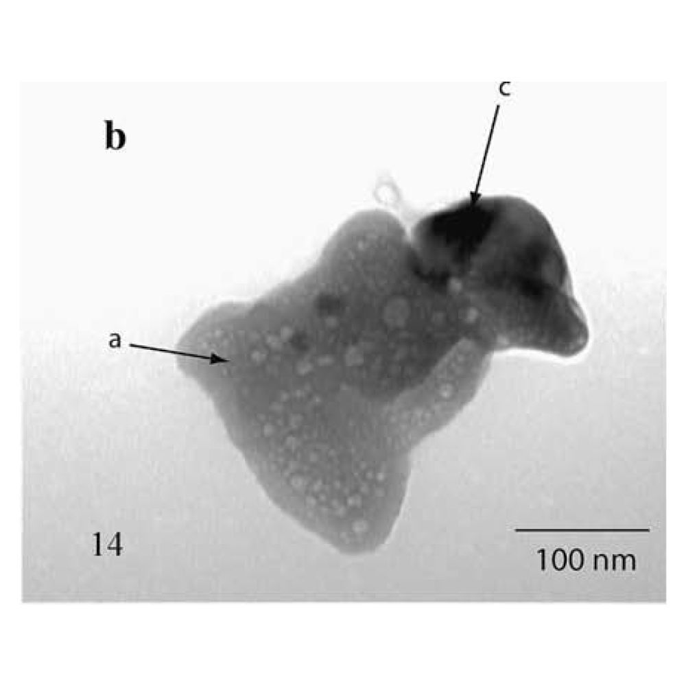
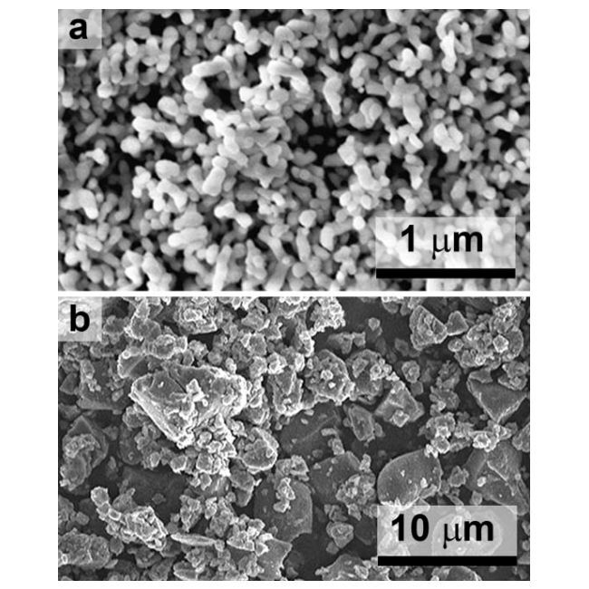
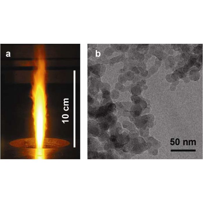
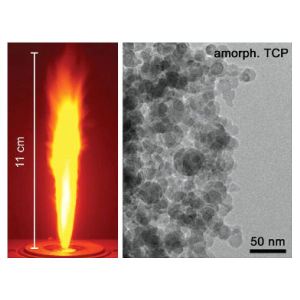
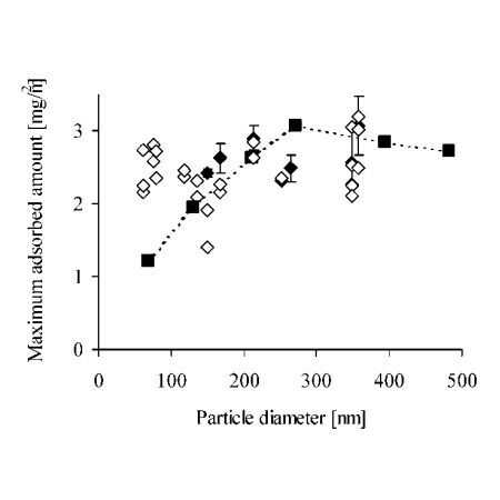
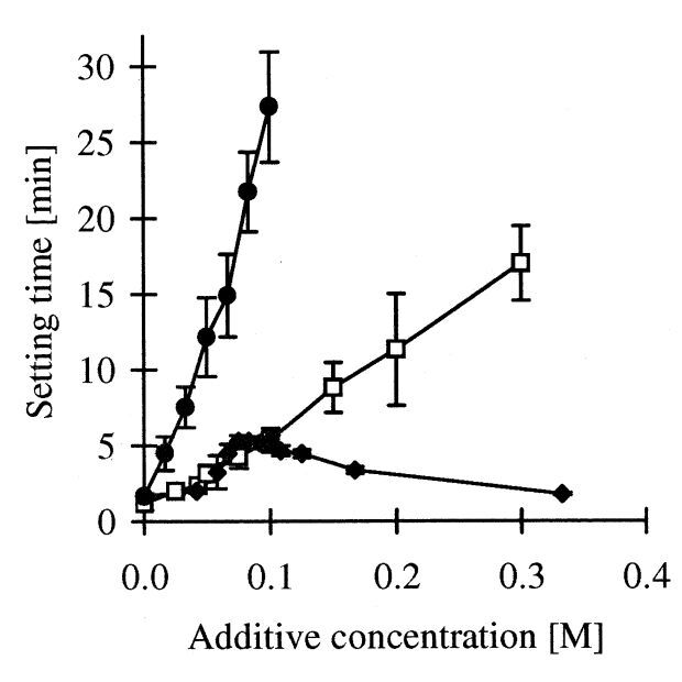
Flame-spray synthesis was used to produce calcium phosphate nanoparticles with a Ca/P molar ratio in the range of 0.5 to 2.0. The use of these powders as raw material for calcium phosphate cements was then assessed. Various characterization techniques revealed that prepared particles consisted of white spheres, had a diameter in the range of 20–30 nm, and formed loose agglomerates. The composition varied from acidic to basic calcium phosphates when the Ca/P molar ratio was increased from 0.5 to 2.0. At the lowest ratio, monocalcium phosphate monohydrate (MCPM) and dicalcium phosphate dihydrate (DCPD) were detected, whereas hydroxyapatite was found at the highest Ca/P molar ratio. All but one of the powders (Ca/P molar ratio = 2.0) reacted with water to form a more crystalline phase. The reaction was highly exothermic and proceeded within a few hours. The reaction product ranged from MCPM/DCPD to hydroxyapatite depending on the initial Ca/P molar ratio. These results suggest that new calcium phosphate cements can be attained using these powders produced by flame-spray synthesis.
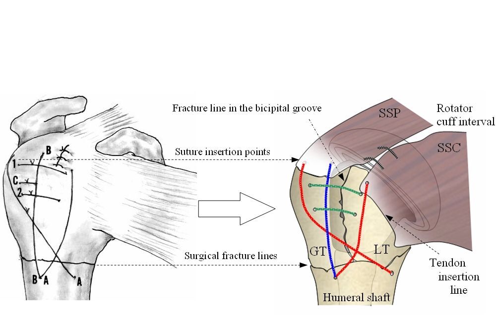
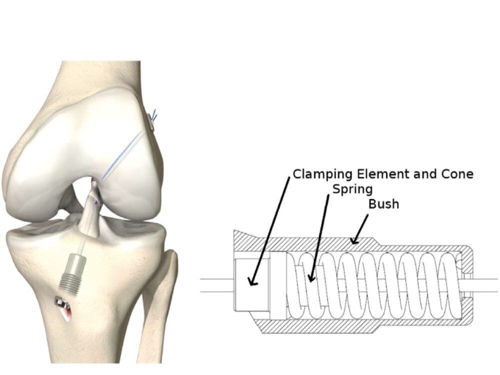
Introduction: The clinical outcome of hemiarthroplasty for proximal humeral fractures is not satisfactory.Secondary fragment dislocation may prevent bone integration; the primary stability by a fixation technique is therefore needed to accomplish tuberosity healing. Present technical comparison of surgical fixation techniques reveals the state-of-the-art approach and highlights promising techniques for enhanced stability.
Method: A classification of available fixation techniques for three- and four part fractures was done. The placement of sutures and cables was described on the basis of anatomical landmarks such as the rotator cuff tendon
insertions, the bicipital groove and the surgical neck. Groups with similar properties were categorized.
Results: Materials used for fragment fixation include heavy braided sutures and/or metallic cables, which are passed through drilling holes in the bone fragments. The classification resulted in four distinct groups: A: both tuberosities and shaft are fixed together by one suture, B: single tuberosities are independently connected to the shaft and among each other, C: metallic cables are used in addition to the sutures and D: the fragments are connected by short stitches, close to the fragment borderlines.
Conclusions: A plurality of techniques for the reconstruction of a fractured proximal humerus is found. The categorisation into similar strategies provides a broad overview of present techniques and supports a further development of optimized techniques. Prospective studies are necessary to correlate the technique with the clinical outcome.
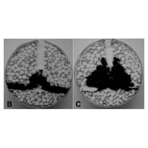
Study Design.
Experimental design using a laboratory leakage model.
Objective.
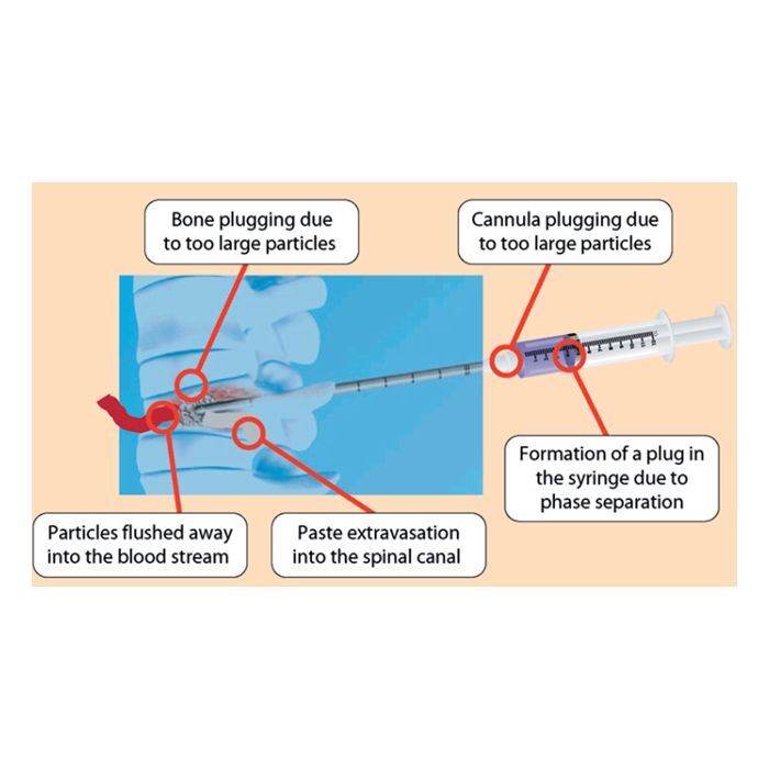
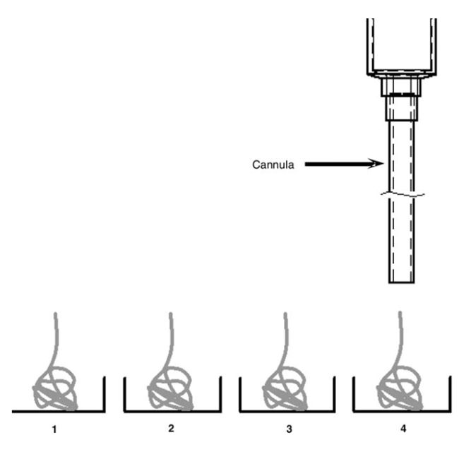
To examine that a new aspiration technique, with a double conduit cannula design, improves the uniformity of cement filling, thus significantly reducing the risk of extraosseous leakage.
Summary of Background Data.
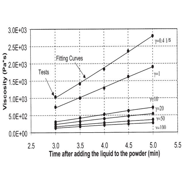
In vertebral augmentation, understanding the forces governing the intravertebral cement flow is essential for controlling the cement formation. A path of least resistance posed by the irregularities in the bone matrix or vertebral shell increases the risk of leak. We have previously shown that using viscous cement reduces the leakage risk. However, this may damage the already weak bone due to the high forces required for the cement to enter the bone cavities.
Methods.
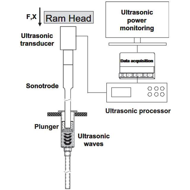
An experimental leakage model for vertebral augmentation was used—with a path, simulating a blood vessel, to provoke leakage. A novel cannula with 2 concentric conduits was used. The inner conduit is used for cement delivery and the outer conduit for aspiration. A mixed level with 2 factors (2 × 22) experiment design was used to examine the ability of aspiration to direct the cement flow in both low and high viscous cement regimes.
Results.
Aspiration significantly enhanced the filling uniformity and reduced the risk of leakage. The reduction in leak with the suction simultaneous to the injection for low viscosity cement, elapsed time 4 minutes, was 1.5 cc (α = 0.05). In the suction experiments, the reduction in leakage as compared with the reference condition for the 8 minutes elapsed time was 0.5 cc, (α = 0.05).
Conclusion.
The aspiration technique combined with a new cannula design improved the uniformity of filling. The aspiration technique helps in removal of the displaced bone marrow or tumor tissue. The aspiration applied with the new cannula requires only a single incision. Thus, it does not result in an increased invasiveness.


Objective
Single-nucleotide polymorphism (SNP) rs143383 (T to C) in the 5′-untranslated region (5′-UTR) of GDF5 has recently been reported to be associated with osteoarthritis (OA) susceptibility, with lower expression of the risk-associated T allele observed in vitro and in vivo. The in vivo studies were performed on cartilage tissue from OA patients. The present study was undertaken to expand the analysis of the effect of this SNP on GDF5 allelic expression to more joint tissue types, to investigate for cis and trans factors that interact with the SNP, and to examine novel cis-acting GDF5 regulatory polymorphisms.
Methods
Tissue samples were collected from OA patients undergoing joint replacement of the hip or knee. Nucleic acid was extracted, and, using rs143383 and an assay that discriminates and quantifies allelic expression, the relative amount of GDF5 expression from the T and C alleles was measured. Additional common variants in the GDF5 transcript sequence were interrogated as potential regulatory elements using allelic expression and luciferase reporter assays, and electrophoretic mobility shift assays were used to search for trans factors binding to rs143383.
Results
We observed a consistent allelic expression imbalance of GDF5 in all tissues tested, implying that the functional effect mediated by rs143383 on GDF5 expression is joint-wide. We identified a second polymorphism, located in the 3′-UTR of GDF5, that influenced allelic expression of the gene independent of rs143383. Finally, we observed differential binding of deformed epidermal autoregulatory factor 1 (DEAF-1) to the 2 alleles of rs143383.
Conclusion
These findings show that the OA susceptibility mediated by polymorphism in GDF5 is not restricted to cartilage, emphasizing the need to consider the disease as involving the whole joint. The existence of an additional cis-acting regulatory polymorphism highlights the complexity of the regulation of expression of this important OA susceptibility locus. DEAF-1 is a trans-acting factor that merits further investigation as a potential tool for modulating GDF5 expression.


Background
Analogous to vertebroplasty, cement-augmentation of the proximal femur (“femoroplasty”) could reinforce osteoporotic bones. This study was to evaluate (i) the feasibility of femoroplasty with a composite cement (Cortoss™), (ii) its influence on femoral strength by mechanical testing and (iii) the feasibility of stable osteosynthesis of the augmented fractured bones.
Methods
Nine human cadaveric femora were augmented with a composite bone cement, the surface heat generation monitored, and then tested biomechanically against their native contralateral control to determine fracture strength. Subsequently, thirteen reinforced and fractured femora were osteosynthetized by different implants and tested against their osteosynthetisized, non-augmented contralateral control.
Findings
Cement could be injected easily, with a moderate temperature rise. A positive correlation between BMD and fracture load and a significant increase in fracture load (+43%) of the augmented femora compared to their native controls (6324N and 4430N, respectively) as well as a significant increase in energy-to-failure (+187%, 86Nm and 30Nm, respectively) was found. Osteosynthesis was possible in cement-augmented femora. Osteosynthetisized femora showed equivalent strength to the intact controls.
Interpretation
Augmentation of the proximal femur with composite bone cement could be of use in prophylaxis of fractures in osteoporotic femurs. Osteosynthesis of the fractured augmented bones is a challenging procedure but has a good chance to restore strength.
Background
Crossed k-wire osteosynthesis is a widely used procedure for displaced supracondylar humerus fractures in children, but the rate of secondary displacements is up to 31%. Alternative techniques including casts, elastic stable intramedullary nailing, and the fixateur extern, have been used, but there are no biomechanical data comparing these methods. We developed a biomechanical model to compare four osteosynthesis techniques for stabilizing supracondylar humerus fractures in children.
Methods
An osteotomy to simulate a fracture was made in a total of 32 adult cadaver humeri. The pseudofractures were then stabilized by crossed k-wires, elastic nailing, a fixateur extern with either k-wires, or Schanz screws. We measured the stiffness values in flexion and extension and torsion with static loading. The movements in cyclic loading were chosen to resemble the mechanism described in the development of a clinical cubitus varus.
Findings
No significant differences were found with static loading. With cyclic loading all methods showed an irreversible torsional deformation less than 20°. Crossed k-wires and elastic nailing showed significantly lower reversible torsional deformation than the external fixateurs.
Interpretation
Our biomechanical data reveal that the crossed k-wires have the highest stiffness and lowest loss of reduction under cyclic loading. The external fixators proved to be good alternatives.
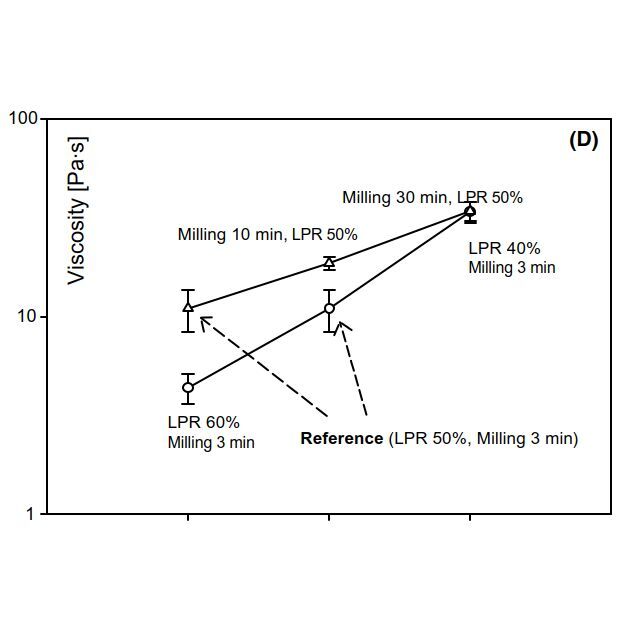
Study Design.
Experimental study using a laboratory leakage model.
Objective.
To examine the working hypothesis that high-viscosity cements will spread uniformly, thus significantly reducing the risk of leakage.
Summary of Background Data.
In vertebroplasty, forces that govern the flow of bone cement in the trabecular bone skeleton are an essential determinant of the uniformity of cement filling. Extraosseous cement leakage has been reported to be a major complication of this procedure. Leakage occurs due to the presence of a path of least resistance caused by irregularities in the trabecular bone or shell structure. Ideally, cement uniformly infiltrates the trabecular bone skeleton and does not favor specific paths. Cement viscosity is believed to affect the infiltration forces and flow during the procedure. Clinically, altering the time between cement mixing and delivery modifies the viscosity of bone cement.
Methods.
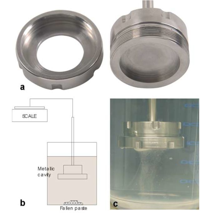
An experimental model of the leakage phenomenon of vertebroplasty was developed. A path, simulating a blood vessel, was created in the model to perturb the forces underlying cement flow and to favor leakage. Cement of varying viscosities was injected in the model, and, thereafter, the filling pattern, cement mass that has leaked, time at which leakage occurred, and injection pressure were measured.
Results.
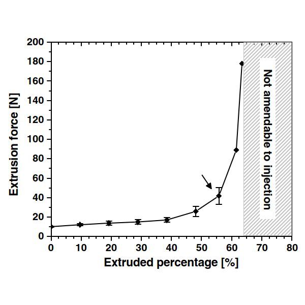
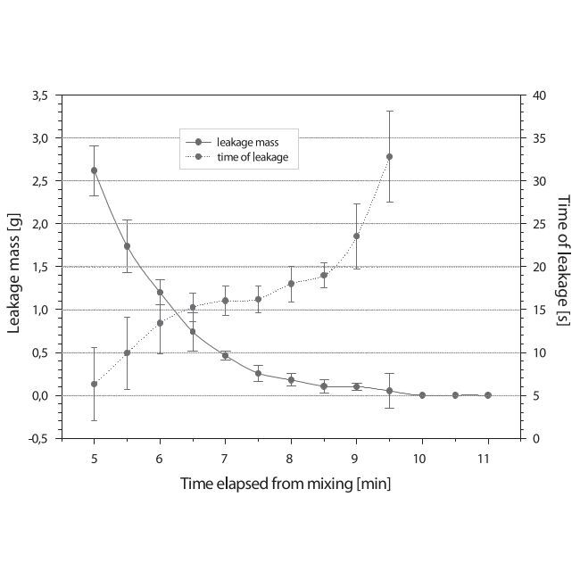
A strong relationship was found between the uniformity of the filling pattern and the elapsed time from cement mixing and viscosity, respectively. Specifically, 3 distinct cement leakage patterns were observed: immediate leakage was observed when cement was injected 5–7 minutes following mixing. The cement was of a low viscosity and more than 50% of the total cement injected leaked. Moderate leakage was observed when injection occurred 7–10 minutes following mixing. Less than 10% of the cement leaked, and the viscosity was at a transient state between the low viscosity of immediate leakage and a higher viscosity, doughy cement. Cement leakage ceased completely when cement was delivered after 10 minutes. The viscosity of the cement in this case was high, and the cement was of a dough-like consistency.
Conclusions.
High-viscosity cement seems to stabilize cement flow. However, the forces required for the delivery of high-viscosity cement may approach or exceed the human physical limit of injection forces. Although the working time of the cement is about 17 minutes, it may not be manually injectable with a standard syringe and cannula after 10 minutes, at which time cement leakage ceased completely.
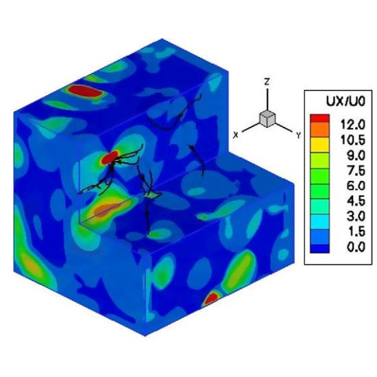
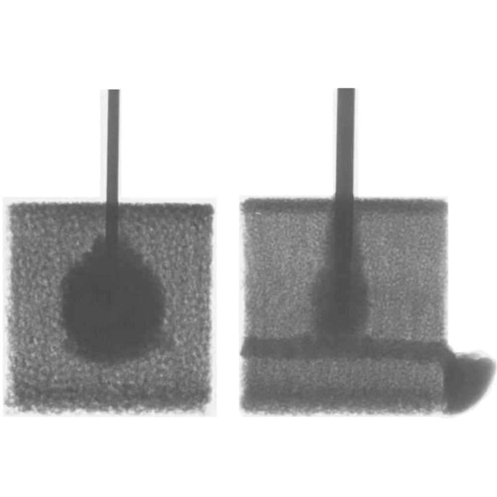
Objectives. – To examine the biomechanisms underlying adjacent fractures following vertebroplasty, an emerging procedure to stabilize fractured vertebrae. In this procedure, bone cement is injected percutaneously into the vertebral cancellous bone. Once hardened, the cement offers mechanical reinforcement to the weakened vertebra. Recent clinical and biomechanical reports suggest that this procedure may cause new fractures adjacent to the one augmented. The cause and extend is unclear yet. The focus here is on the biomechanical hypothesis resulting from the rigid cement augmentation.
Methods. – A combination of experimental and numerical studies, in additional to a review of recent clinical reports.
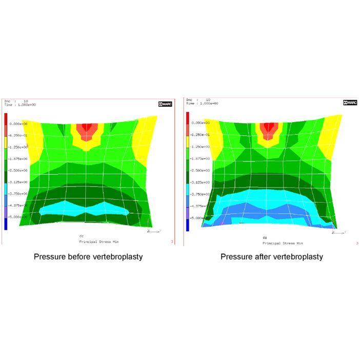
Results. – The broader finding suggests that vertebroplasty changes the mechanical loading in adjacent vertebrae. Specifically, an increase in adjacent loading in the range of 17% has been found. The mechanism underlying this increase seemed to stem from the excessive cement rigidity that reduced the endplate bulge of the augmented vertebra, thereby reducing the local spinal joint flexibility. The reduction in joint flexibility seeks to reverse itself by creating an increase in the inter-vertebral disc pressure. The increased disc pressure seeks to relieve itself by increasing the load on the adjacent vertebra. The increased load on the adjacent vertebra relates directly to an increased risk of fracture.
Conclusions. – Although an increasing amount of evidence exists to support this theory of the origin of adjacent fractures, one must be cautious. Vertebroplasty is a relatively new procedure and further observations and, ultimately, prospective clinical studies are required to conclusively determine the cause and extend of adjacent fractures.
Objectifs. – Examiner les biomécanismes sous-jacents aux fractures apparaissant suite à la vertébroplastie - une procédure émergente visant à stabiliser les vertèbres fracturées. Lors de cette procédure, un ciment osseux est injecté par voie percutanée au travers de l’os spongieux vertébral. Une fois durci, le ciment offre un renforcement mécanique à la vertèbre affaiblie. Des études biomécaniques et cliniques suggèrent que cette procédure puisse induire des fractures adjacentes à celle consolidée. La cause et son importance ne sont actuellement pas claires. L’étude se concentre sur l’hypothèse biomécanique résultante de la rigidité imposée lors de la consolidation par ciment.
Méthodes. – Une combinaison d’études expérimentales et numériques a été menée, en plus d’une revue littéraire d’articles cliniques récents.
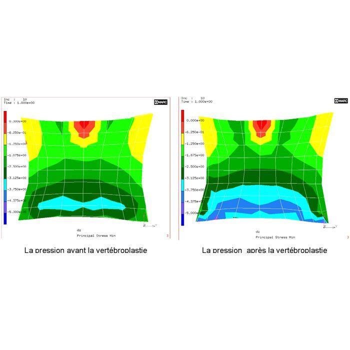
Résultats. – Les résultats généraux suggèrent que la vertébroplastie change les chargements mécaniques appliqués sur les vertèbres adjacentes. Plus spécifiquement, une augmentation de ces chargements de 17% a été reportée. Le mécanisme sous-jacent à cette augmentation semble provenir de l’extrême rigidité du ciment, réduisant la flexibilité de la plaque supérieure de la vertèbre consolidée, ainsi réduisant la flexibilité du joint intervertébral. La réduction de la flexibilité de ce joint est alors compensée par une augmentation de la pression du disque avoisinant. L’augmentation de la pression de ce disque est de même compensée par une augmentation de la charge sur la vertèbre adjacente. Cette augmentation de la charge sur la vertèbre adjacente est alors directement liée à une augmentation du risque de fracture.
Conclusions. – Malgré le fait qu’il existe de nombreuses études cliniques supportant cette hypothèse des fractures adjacentes, il nous faut être prudent. La vertébroplastie est une procédure relativement nouvelle et de plus amples observations, et ultimement, des études cliniques prospectives sont requises afin de conclure sur la cause et l’importance des fractures adjacentes.


Objective:
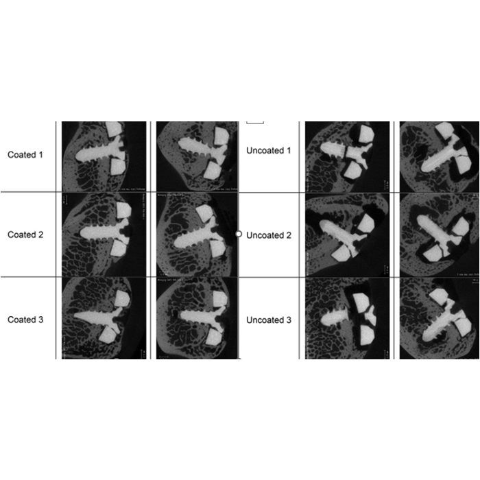
This study was designed to assess the benefits of a new Anodic Plasma Chemical calcium-phosphate (APC-CaP) surface treatment on reducing pin track infection and pin loosening in comparison to anodized titanium (Ti) during external fracture fixation.
Methods:
A tibial midshaft, transverse, 6-mm gap osteotomy was created in 17 adult female Swiss alpine sheep. The tibia was stabilized with an external fixator and 4 Schanz screws of Ti or APC-CaP-treated Ti. The sheep were examined during a 12-week observation period. Infection was assessed with weekly clinical pin track grading and microbiologic assessment at sacrifice. Pin loosening was assessed by grading for radiolucency on biweekly radiographs and by measuring extraction torque on pin removal. In vivo bending stiffness measurements were performed to determine gap healing. A qualitative histologic assessment of the tissue adjacent to pin sites was also performed.
Results:
A trend (P = 0.056) for less infection around APC-CaP pins was found at 6 weeks, but the strength of this difference diminished with time. Significantly more radiolucency was found around Ti pins after 8 (P = 0.011) and 12 (P < 0.001) weeks. At all pin sites, the extraction torque for APC-CaP pins was higher than for Ti pins (P = 0.007). No difference in the progression of gap healing was found. Histology showed bone growth at the implant surface in the form of distance osteogenesis for Ti and contact osteogenesis for APC-CaP.
Conclusions:
This study has shown that the APC-CaP surface improves the clinical performance of Ti pins with respect to pin loosening and pin track infection.
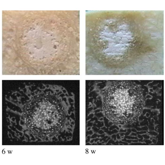
Introduction
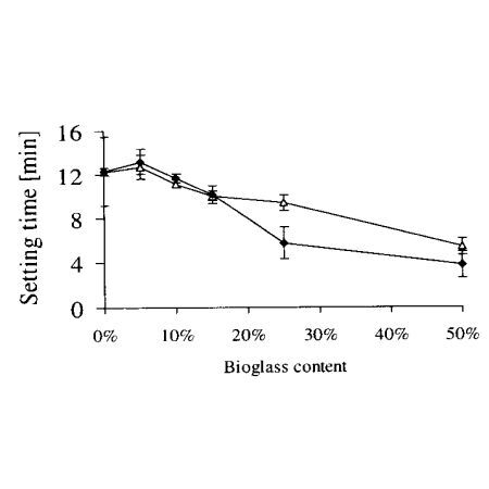
Aim of this experimental study was to assess the suitability of a new brushite calcium phosphate cement (chronOS™Inject) for cranioplasty and to compare the results with a commercially available apatite calcium phosphate bone cement (Biobon®).
Material and methods
A bilateral full-size craniotomy defect (23mm in diameter) was created in the parietal bones of 18 adult Swiss Alpine sheep and filled with either chronOS™Inject or Biobon®. The observation intervals were 2, 4 and 6 months. Macroscopical, radiological, histological and histomorphometrical evaluations were performed.
Results
New bone formation was moderate and did not differ significantly between the biomaterials. Cement resorption occurred centripetally in the chronOS™Inject group and proceeded significantly faster than the degradation process of Biobon®. However, implantation of chronOS™Inject was associated with a significantly higher rate of fibrous tissue formation. Cement resorption was mediated by macrophages in the chronOS™Inject group, while osteoclasts were the predominant cell type involved in degradation of Biobon®. Osteoblasts were found adjacent to residual cement in both groups.
Conclusion
chronOS™Inject demonstrated osteoconductive properties, good biocompatibility and superior bioresorbability but none of the cements proved suitable for filling large cranial bony defects due to the high rate of fibrous tissue formation and insufficient bony regeneration.

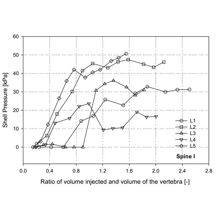
Study Design.
An experimental biomechanical study conducted on osteoporotic cadaveric vertebrae.
Objectives.
1) To measure the intravertebral shell pressure and injection pressure; and 2) to determine the effect of the vertebral shell on the intravertebral shell pressure and on the injection pressure.
Summary of Background Data.
Forces that govern cement flow are an essential component of the cement injection process in vertebroplasty. The vertebral shell may play a significant role in confining the flow of cement in the vertebral body and thereby affecting the intravertebral pressure and injection pressure.
Methods.
A small fenestration was created in the left lateral vertebral shell of 14 vertebrae. A valve to open and close the fenestration and a sensor to measure the intravertebral pressure were attached to the opening. A closed fenestration simulated an intact shell, whereas an open fenestration represented a vented shell. Injection pressure and intravertebral pressure at the shell were recorded during a controlled injection.
Results.
A closed fenestration resulted in a significant increase in the intravertebral pressure at the shell. During the injection, the shell pressure increased on average to approximately 3.54 ± 2.91 kPa. Conversely, an open fenestration resulted in an instant relaxation of the shell pressure to the ambient pressure of 0 kPa. Additionally, the injection pressure was approximately 97 times higher than the shell pressure.
Conclusion.
The presence of vertebral shell seems to be important for intravertebral pressure. However, the intravertebral shell pressure adds very little to the injection pressure.
Objective:
To compare the vertical subsidence in a bicondylar tibial plateau fracture model stabilized either by a unilateral locked screw plate (LSP) or by double plating.
Design:
Biomechanical cadaver study.
Intervention:
A 41-C1 fracture model was created in eight pairs of fresh-frozen human cadaver tibiae. Stabilization was performed either by open reduction and internal fixation (ORIF) using a lateral L-buttress plate and a medial four-hole, one-third tubular antiglide-plate or by a lateral LSP. Four load levels (400N, 800N, 1200N, 1600N), each with five cycles, were consecutively applied to the medial plateau.
Main Outcome Measurements:
The vertical plastic deformation at the end of each cycle was the main parameter of interest. Statistical analysis was performed with the two-way ANOVA test for repeated measurements. Each individual loading level was analyzed separately using Student t test.
Results:
In one pair, both fixation techniques failed at the first loading cycle of 1200N. One ORIF fixation failed at the first loading cycle of 1600N. The average plastic vertical subsidence was 0.40 mm (LSP) and 0.25 mm (ORIF) at 400N (P = 0.291), 0.83 mm (LSP) and 0.81 mm (ORIF) at 800N (P = 8.82), 1.06 mm (LSP) and 0.96 mm (ORIF) at 1200N (P = 0.98), and 1.54 mm (LSP) and 1.14 mm (ORIF) at 1600N (P = 0.53). Vertical subsidence depended on the applied load (P = 0.002), but not on the method of fixation (P = 0.236).
Conclusion:
Both fixation techniques have a high resistance to vertical subsidence even with loads exceeding the average body weight. No statistically significant difference was seen between the two methods of fixation.
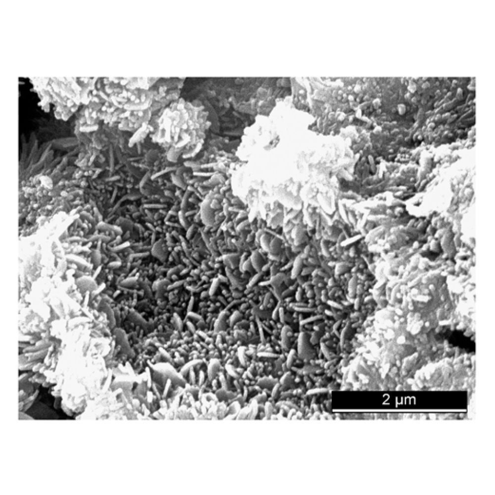
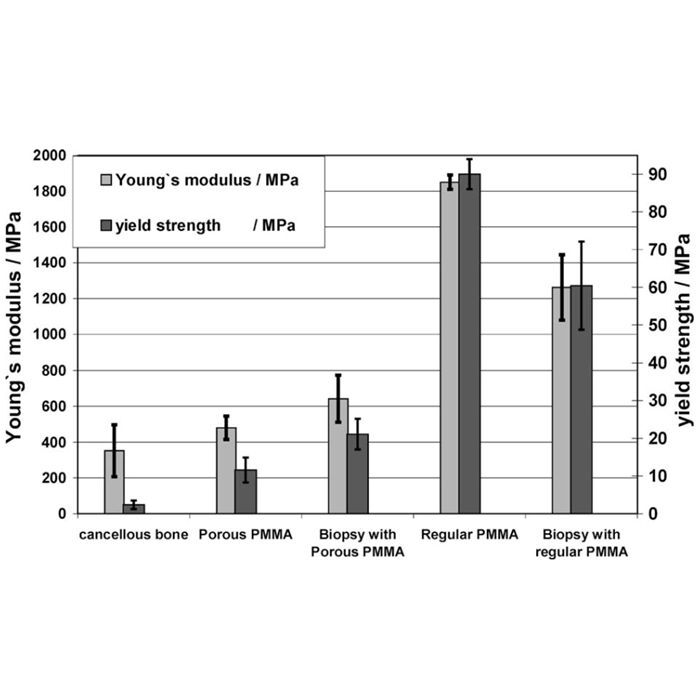
Objective. To determine the feasibility of polymethyl-methacrylate injection into the osteoporotic proximal femur and its effect on the mechanical properties.
Design. In vitro pairwise comparison of non reinforced and reinforced bones in a load to failure loading mode.
Background. Hip fractures represent an important public healthcare problem. Continued growth in the elderly population will raise the incidence of hip fractures and their associated costs dramatically in the near future.
Methods. Twenty pairs of osteoporotic femurs were mechanically tested either in a single-limb stance configuration or simulating a fall on the greater trochanter. From each pair, one femur was augmented with bone cement, with the contralateral femur serving as a control. The surface temperature at the femoral neck was recorded until twenty minutes after injection. The fracture load and the energy absorption were calculated. The Wilcoxon signed rank test was used to test for differences in fracture load and energy absorption between the reinforced femurs and the native controls.
Results. Volumes of 28–41 ml of cement (mean, 36 ml) could be injected. The increase of surface temperature at the femoral neck ranged from Δ18.4 to Δ29.8 °C. For the single limb stance configurations, the peak fracture load was increased by 21%, (P<0.002) and for the simulated fall on the hip by 82%, (P<0.002). The corresponding values for energy absorption were +48%; and +188% (P<0.002) respectively.
Conclusions. The feasibility and mechanical effectiveness of the in vitro procedure could be demonstrated. The heat generation due to polymethyl-methacrylate polymerisation is high.
Relevance Prophylactic reinforcement of the femur could become a treatment option to solve the problems with osteoporotic hip fractures in patients at risk. Reinforcement materials with less exothermic reaction need to be evaluated further and also the feasibility of fracture repair after reinforcement.
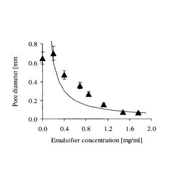
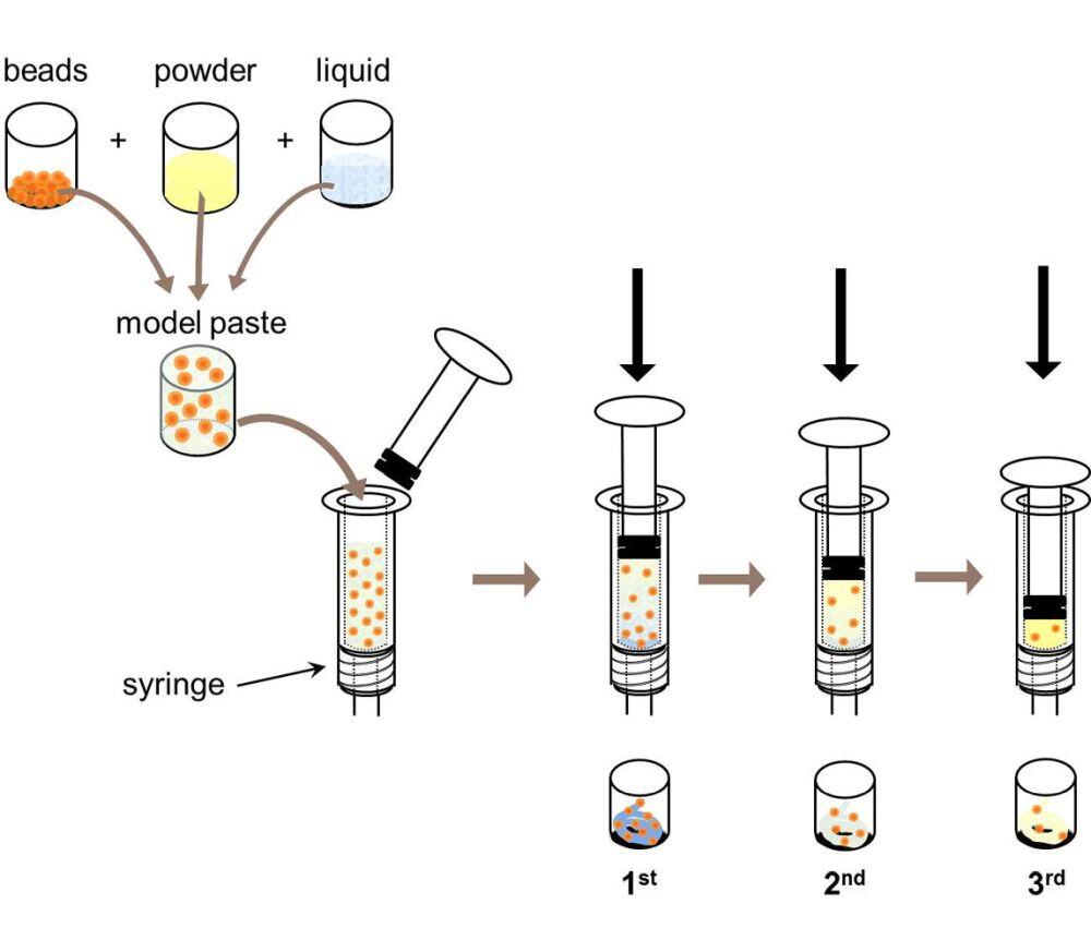
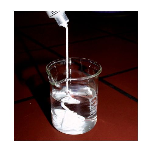
A hydrophobic liquid was mixed with a calcium phosphate cement paste and an emulsifier. After intensive mixing, an emulsion was formed. The emulsion was either a W/O emulsion (cement droplets dispersed in oil) or a O/W emulsion (oil droplets dispersed in the cement paste), depending on the emulsifier type and the oil stability. The stability of the two types of emulsion could be well predicted using the hydrophilic-lipophilic balance concept. After the cement had set and the oil had been washed off, calcium phosphate granules or porous blocks were obtained. The composition of the granules and the blocks was either calcium-deficient hydroxyapatite (before sintering) or β-tricalcium phosphate (after sintering). An increase of the emulsifier concentration or a decrease of the oil amount led to a decrease of the block pore size.
Objective
Comparison between a Less Invasive Stabilization System (LISS) using monocortical screws with angular stability and two conventional plate systems Condylar Buttress Plate (CBP) and Dynamic Condylar Screw (DCS) for the treatment of distal femoral fractures with respect to biomechanical properties.
Design
Biomechanical study using paired cadaver femurs. In Test Configuration 1 (distal test), a ten-millimeter gap at the diaphysis–metaphysis junction simulates a supracondylar femoral fracture. Test Configuration 2 (proximal test) has the same configuration, but the gap was cut in the isthmic region. Proximal and distal plate ends were fixed to corresponding cortical bone fragments in both tests. Optical displacement transducers served to quantify the system's ability to withstand a stepwise increased load. Reversible (deflection) and irreversible deformation (subsidence) of the bone–plate construct was investigated.
Results
In Test Configuration 1, LISS showed less irreversible deformation in 72 percent of the left–right comparisons. No correlation between bone mineral density, cross-section area of bones and the measured response of the construct under load was found between pairs. In Test Configuration 2, 83 percent of the left–right comparisons showed less permanent deformation but a higher elastic deformation for LISS.
Conclusions
These results suggest an enhanced ability to withstand high loads when using the monocortical screw fixation technique with angular stability. A higher elastic deformation of LISS compared with conventional plating systems in distal femoral fractures can be explained by the lower bending stiffness caused by different design and material properties.

Die Dienstleistungen unseres Materialprüflabors sind seit 1995 nach ISO/IEC 17025 akkreditiert. Unser QM-System ist ISO 9001 zertifiziert.


Hier finden Sie unsere neuesten Blog-Beiträge.
![Save the Date - [MEET THE EXPERT] Implants](https://www.rms-foundation.ch/hubfs/01%20Bilder%20-%20Logos%20-%20Icons/Bilder%20MtE/Banner_MtE.jpg)

RMS Foundation
Robert Mathys-Strasse 1
2544 Bettlach
Schweiz
Abonnieren Sie unseren Info-Letter und wir informieren Sie etwa 10-mal pro Jahr über aktuelle Entwicklungen in den Bereichen Materialprüfung, Forschung und Wissenstransfer.