IL-34: Particle analysis: How to characterise particles
In order to prove the safety of a product, many medical device manufacturers determine the type, quantity and size of the abrasion particles, which are formed in a simulated mechanical stress situation. Such tests are also increasingly required by the regulatory authorities.
Typical laboratory investigations to determine the particle generation on implants (e.g. dental, orthopaedic or trauma implants) first include a loading test. Here the products are subjected to a cyclical alternating load that is as realistic as possible. The mechanical load is usually applied in physiological fluid and at body temperature.
During the subsequent particle collection, the highest level of cleanliness must be ensured in order to avoid additional contamination. For this reason, the work is carried out in a clean room. In order to exclude further side effects of the processes, blind and reference samples are always carried along.
![]()
Figure 1: Dynamic loading of a dental implant to generate particles.
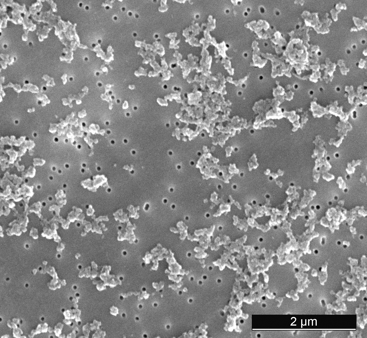
The number and size of the wear particles in liquids can be determined by light obscuration. For a more detailed analysis the particles are collected on a filter. The quantity of the generated particles is determined gravimetrically by comparing the mass of the loaded and conditioned filter with that of the filter before the particle loading. The filters are then evaluated by automated light microscopy with regard to the number, size, shape and type of particles (see Newsletter 28). Small particles that are not visible with light microscopes are examined with a scanning electron microscope (SEM). In combination with an EDX analysis, the chemical composition can also be determined.
The results of the particle analyses serve as a basis for the evaluation of the safety of a medical product under the selected application conditions.

Figure 2: SEM image of particles on a filter (top) and colour coding of the individually analysed particles (bottom).
Our possibilities for particle collection and analysis on surfaces of medical devices:
- Implant specific dynamic tests
- Ultrasonic bath Bandelin Sonorex Digiplus DL512H
- Light obscuration AccuSizer A7000 SIS
- Vacuum filtration unit Sartorius
- Analytical balance Mettler XPE 2015
- Filter analysis system Jomesa HFD4
- Scanning electron microscope Zeiss Sigma 300 VP with EDX-analysis and software AZtecFeature
- FT-IR microscope Bruker Lumos
- ICP-MS Agilent 7700x
