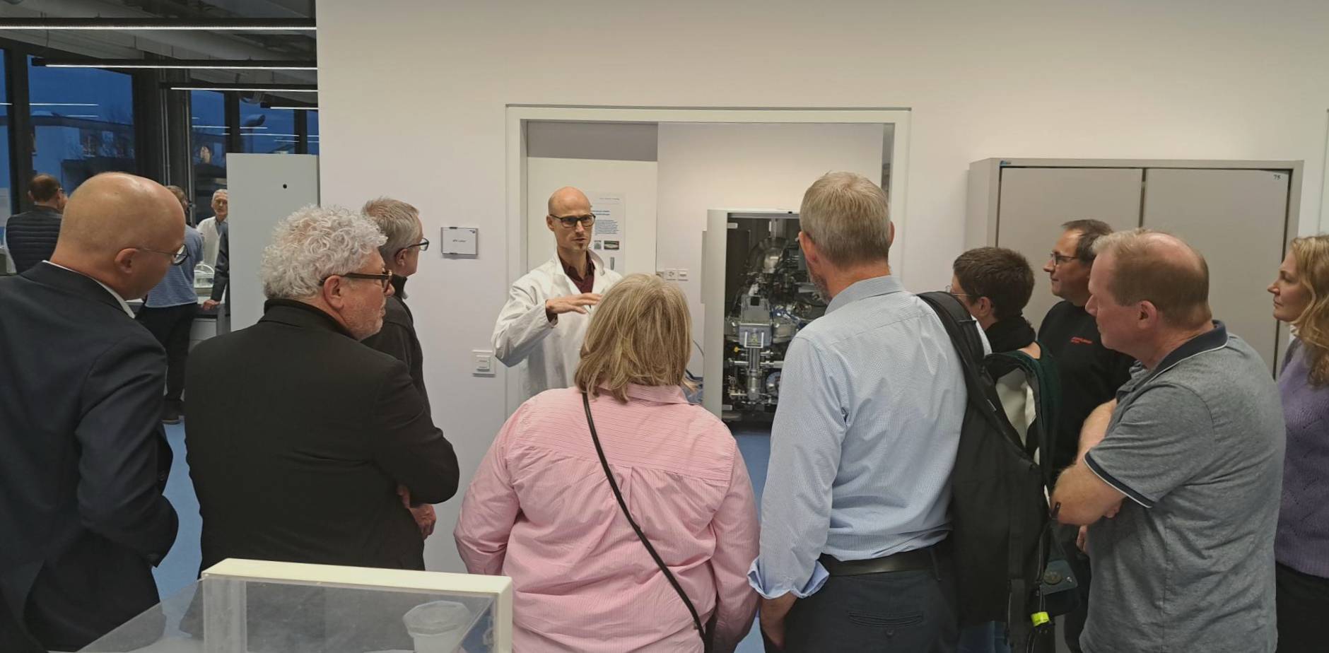
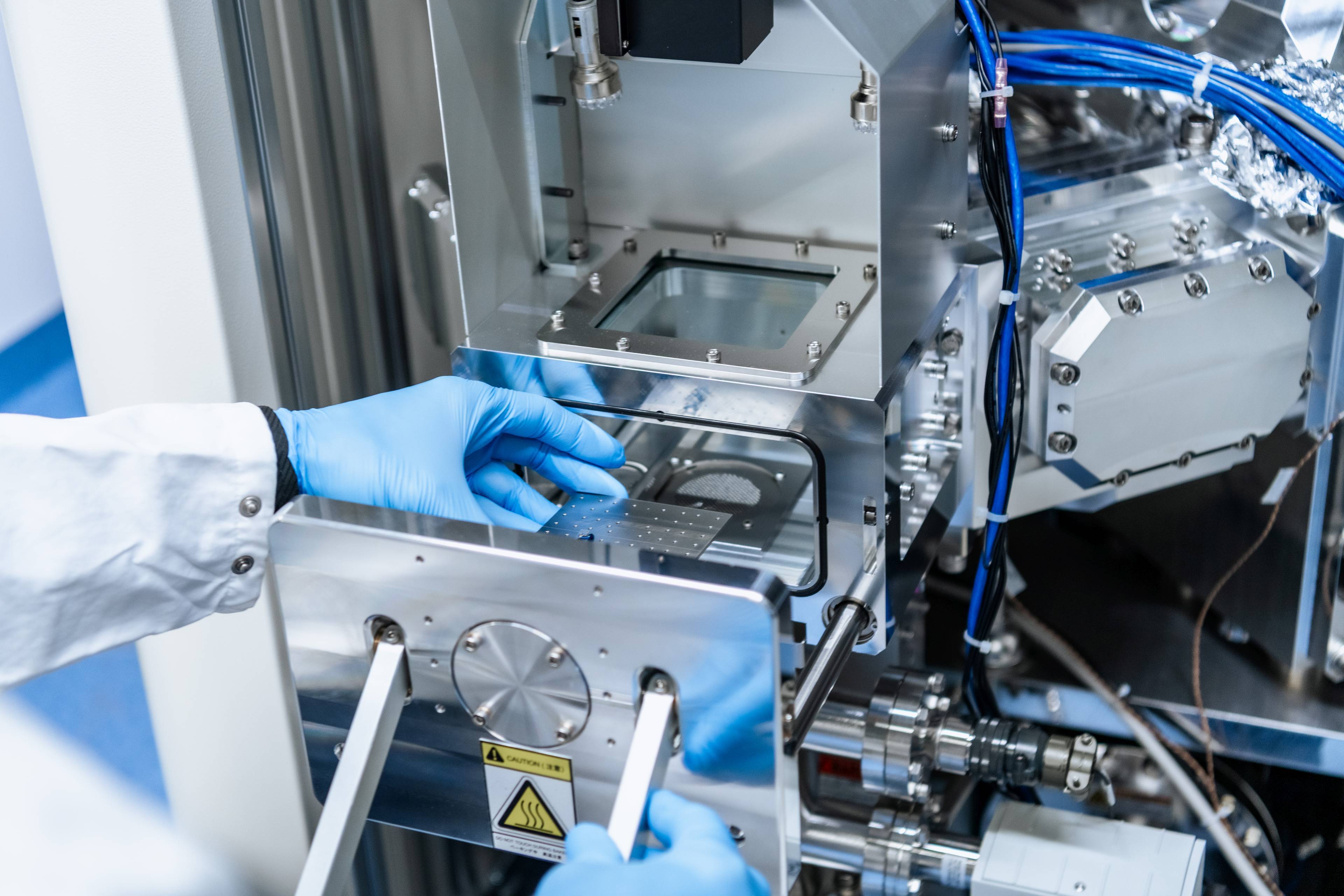
The FTIR microscope can be operated in the three measuring modes; ATR (Attenuated Total Reflection), transmission, and reflection. For the investigation of larger samples an additional conventional FTIR spectrometer (Macro Unit) is connected to the microscope (Figure 1). The chemical composition of inhomogeneous structures can be visualized by mapping the sample through stage motorization in the x/y/z directions.
 Figure 1: Bruker LUMOS FTIR microscope for measurements in transmission, reflection and ATR, coupled with a Macro Unit for large samples.
Example 1: Ultrahigh molecular weight polyethylene (UHMWPE) has been used for many years in orthopedics for acetabular components. However irradiated UHMWPE has a poor long-term resistance to oxidation when in the presence of oxygen. By adding small amounts of vitamin E as an antioxidant, the free radicals are saturated. This stabilizes the material by preventing oxidative damage. Figure 2 shows an oxidation index depth profile - collected according to ASTM F2102 - of an explant from conventional UHMWPE (non-stabilized) with strong oxidation of the material. The oxidation index is the ratio of the absorbance peaks at 1720 cm-1 normalized to the reference peak at 1370 cm-1. The greatest damage occurred at a depth of 0.4 mm, since the compressive stresses are greatest below the articular surface.

Figure 2: Oxidation index depth profile measured on an implant made of UHMWPE.
Example 2: Unknown fibers on the surface of an implant were identified in the FTIR microscope as nylon (polyamide 66, Figure 3). It was then possible to determine their origin and to solve the problem of contamination.

Figure 3: Image of the measurement surface and corresponding FTIR spectrum of a nylon fiber.
Equipment:
FTIR microscope Bruker LUMOS
Analyisis options:
- PMI analysis (Positive Material Identification) of organic material (bulk solids, powders, liquids) in accordance with ASTM F1252.
- Investigation of layered structures such as: packaging, polymer laminates, and chips in paint and lacquer.
- Identification of small organic particles, fibers, or contamination on surfaces.
- Quantitative component analysis of organic substances.
- Oxidation Index and Trans-Vinylene Yield as determined by ASTM F2102 / F2381.

![Save the date - [MEET THE EXPERT] Implants](https://www.rms-foundation.ch/hubfs/01%20Bilder%20-%20Logos%20-%20Icons/Bilder%20MtE/Banner_MtE.jpg)