In artificial joint replacements for example, the roughness of articulating surfaces strongly influences the wear which is generated in these devices. In the case of dental and orthopaedic implants, the material and its surface structure particularly determine bone ongrowth and therefore its fixation to bone.
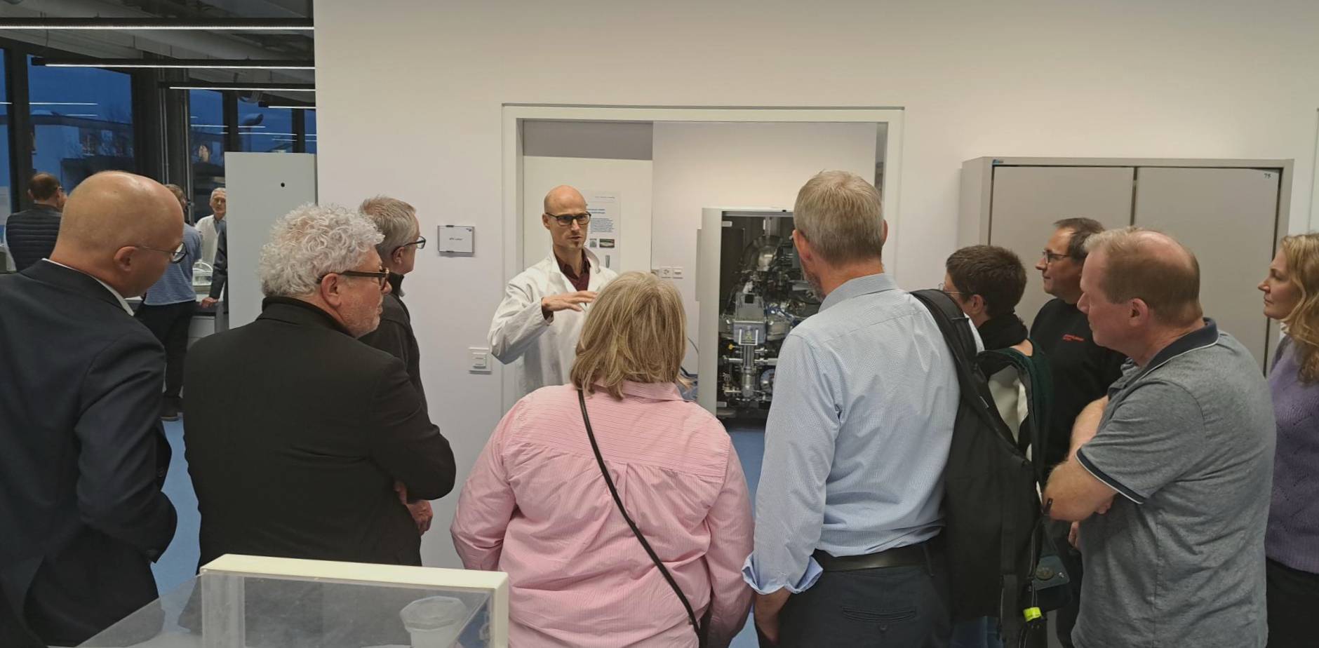
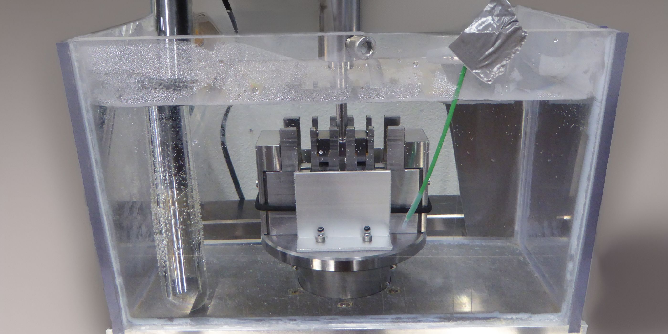
At the RMS Foundation, we use a modern, optical 3D-measuring device to measure the surface structure and roughness on many kinds of samples. The S neox from Sensofar allows measurements in different modes: interferometry (both phase shift and white light interferometry), confocal microscopy and focus variation. With interferometry – combined with a piezo drive and an active vibration control – depth-resolutions in the sub-nanometre range are possible. Rough samples can be investigated using confocal microscopy and the contour of a sample can quickly be assessed with focus variation. Very large samples can be analysed too, thanks to a one metre high column for the sensor head.
If the lateral resolution of optical systems is not sufficient, then the topography can be assessed by scanning electron microscopy (SEM): Pictures of a surface are acquired at two different tilting angles and thus the topography can be determined semi-quantitatively. Herewith lateral resolutions down to few nanometres can be achieved.

Figure 1: Topography of a scratched (3rd-body wear) polymer implant after explantation.
Besides the surface roughness parameters, the more common profile roughness values can be determined by extracting and evaluating multiple profiles from the measured topographies. Several single topographies are usually stitched together to obtain a larger topography, based on which the roughness values can be determined according to ISO standard 21920-3. This can be performed for highly polished surfaces as well as for rough samples.
The S neox is used for various projects, both for testing and research. For example, an artificial ceramic-on-polymer joint was investigated, which was heavily damaged in vivo by 3rd-body wear caused by metallic particles, which entered into the articulation. The surfaces were documented using white light interferometry. It was shown that the softer polymer part was heavily scratched (Figure 1) while there was material transferred onto the ceramic part (Figure 2). This material caused a higher roughness which in turn led to more abrasion of the polymer part.

Figure 2: Topography of a ceramic implant: explant with metal transfer.
Optical roughness measurement
Equipment:
S neox frm Sensofar (Spain)

Measuring modes
• Interferometry (Phase shift and white light interferometry)
• Confocal microscopy
• Focus variation
Resolution
vertical: 0.1 nm with phase shift interferometry
lateral: 0.2 µm (100X objective)
Sample requirements
Solid samples
Dimension: < 60 x 60 x 60 cm
Weight: < 10 kg
Reflectivity: 0.05 % to 100 %
Height difference: < 40 mm
Lateral movement: < 114 x 75 mm
Normative references
Profiles: ISO 21920-1, -2 & -3
Areas: ISO 25178-1, -2 & -3


![Save the date - [MEET THE EXPERT] Implants](https://www.rms-foundation.ch/hubfs/01%20Bilder%20-%20Logos%20-%20Icons/Bilder%20MtE/Banner_MtE.jpg)